• 文献检索
  • 文档翻译
  • 深度研究
  • 学术资讯
  • Suppr Zotero 插件Zotero 插件
  • 邀请有礼
  • 套餐&价格
  • 历史记录
应用&插件
Suppr Zotero 插件Zotero 插件浏览器插件Mac 客户端Windows 客户端微信小程序
定价
高级版会员购买积分包购买API积分包
服务
文献检索文档翻译深度研究API 文档MCP 服务
关于我们
关于 Suppr公司介绍联系我们用户协议隐私条款
关注我们

Suppr 超能文献

核心技术专利:CN118964589B侵权必究
粤ICP备2023148730 号-1Suppr @ 2026

文献检索

告别复杂PubMed语法,用中文像聊天一样搜索,搜遍4000万医学文献。AI智能推荐,让科研检索更轻松。

立即免费搜索

文件翻译

保留排版,准确专业,支持PDF/Word/PPT等文件格式,支持 12+语言互译。

免费翻译文档

深度研究

AI帮你快速写综述,25分钟生成高质量综述,智能提取关键信息,辅助科研写作。

立即免费体验

以组织病理学为金标准的骨肿瘤X线与磁共振成像相关性研究

X-ray and MRI Correlation of Bone Tumors Using Histopathology As Gold Standard.

作者信息

Azad Hina, Ahmed Aliya, Zafar Ibtesam, Bhutta Muzammil Rasheed, Rabbani Muhammad Ali, Kc Himesh Raj

机构信息

Radiology, Pakistan Institute of Medical Sciences, Islamabad, PAK.

Anatomy, CMH Multan Institute of Medical Sciences, Multan, PAK.

出版信息

Cureus. 2022 Jul 25;14(7):e27262. doi: 10.7759/cureus.27262. eCollection 2022 Jul.

DOI:10.7759/cureus.27262
PMID:36039258
原文链接:https://pmc.ncbi.nlm.nih.gov/articles/PMC9403219/
Abstract

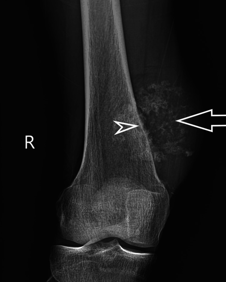
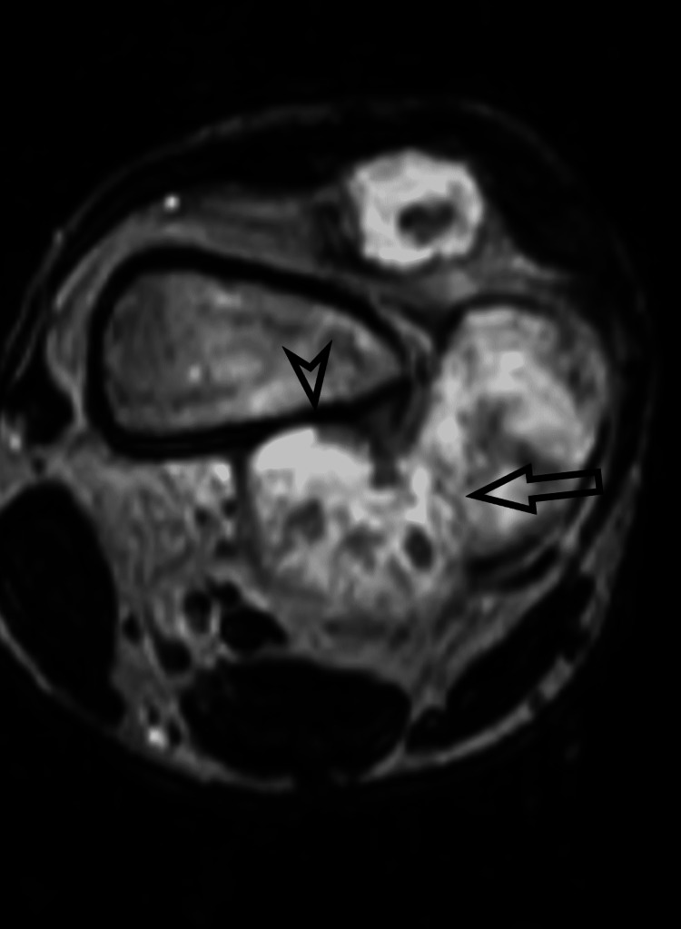
Introduction Bone tumors are a common pathology of the musculoskeletal system being frequently encountered by clinicians. Radiological workup is a mainstay in the diagnostic workup of bone tumors. This study aimed to highlight the importance of plain radiography and MRI in the diagnosis of bone tumors keeping histopathology as a gold standard. It is a descriptive validation study conducted in the Radiology Department of Pakistan Institute of Medical Sciences Islamabad. Methodology The study included 92 patients with suspected bone lesions. After taking a complete history and receiving informed written consent. X-rays radiographs and magnetic resonance imaging were performed. X-ray radiograph and magnetic resonance imaging parameters were recorded and compared with the histopathology of lesions as a standard. Results The mean age of patients was 30.50 ± 8.95 years. Of 92 patients examined on X-ray, 51 (55.4%) had lytic lesions, 34 (37.0%) had sclerotic lesions, and seven (7.6 %) had mixed lesions. MRI revealed the location of the lesion. There were 25 (27.2%) bone lesions in diaphysis, 19 (20.7%) in metaphysis, nine (9.8%) at meta-diaphysis, and 32 (34.8 %) in the meta-epiphyseal region. These findings were later on confirmed with histopathological results. Conclusion MRI can differentiate soft-tissue components and periosteal reactions. An X-ray radiograph can provide information about bony matrix and calcifications within tumors. After analysis of imaging findings and histopathological results, it is concluded that these modalities can be used to diagnose bone tumors with high diagnostic accuracy.

摘要

引言

骨肿瘤是肌肉骨骼系统的常见病理表现,临床医生经常会遇到。影像学检查是骨肿瘤诊断检查的主要手段。本研究旨在强调X线平片和磁共振成像(MRI)在骨肿瘤诊断中的重要性,并将组织病理学作为金标准。这是一项在伊斯兰堡巴基斯坦医学科学研究所放射科进行的描述性验证研究。

方法

该研究纳入了92例疑似骨病变患者。在获取完整病史并获得知情书面同意后,进行了X线平片和磁共振成像检查。记录X线平片和磁共振成像参数,并与病变的组织病理学结果进行比较作为标准。

结果

患者的平均年龄为30.50±8.95岁。在92例接受X线检查的患者中,51例(55.4%)有溶骨性病变,34例(37.0%)有硬化性病变,7例(7.6%)有混合性病变。MRI显示了病变的位置。骨干有25例(27.2%)骨病变,干骺端有19例(20.7%),干骺-骨干交界处有9例(9.8%),骨骺-干骺端区域有32例(34.8%)。这些发现随后通过组织病理学结果得到证实。

结论

MRI可以区分软组织成分和骨膜反应。X线平片可以提供有关肿瘤内骨基质和钙化的信息。在分析影像学表现和组织病理学结果后,得出结论:这些检查方法可用于以高诊断准确性诊断骨肿瘤。

https://cdn.ncbi.nlm.nih.gov/pmc/blobs/bd11/9403219/934eadda8d49/cureus-0014-00000027262-i02.jpg
https://cdn.ncbi.nlm.nih.gov/pmc/blobs/bd11/9403219/4067fd51e501/cureus-0014-00000027262-i01.jpg
https://cdn.ncbi.nlm.nih.gov/pmc/blobs/bd11/9403219/934eadda8d49/cureus-0014-00000027262-i02.jpg
https://cdn.ncbi.nlm.nih.gov/pmc/blobs/bd11/9403219/4067fd51e501/cureus-0014-00000027262-i01.jpg
https://cdn.ncbi.nlm.nih.gov/pmc/blobs/bd11/9403219/934eadda8d49/cureus-0014-00000027262-i02.jpg

相似文献

1
X-ray and MRI Correlation of Bone Tumors Using Histopathology As Gold Standard.以组织病理学为金标准的骨肿瘤X线与磁共振成像相关性研究
Cureus. 2022 Jul 25;14(7):e27262. doi: 10.7759/cureus.27262. eCollection 2022 Jul.
2
The current role of radiography in the assessment of skeletal tumors and tumor-like lesions.放射成像在骨骼肿瘤及肿瘤样病变评估中的当前作用。
Eur J Radiol. 1998 May;27 Suppl 1:S77-85. doi: 10.1016/s0720-048x(98)00047-3.
3
The Performance of Radiographic Criteria for Bone Malignancy When Applied to Computed Tomography and Magnetic Resonance Imaging.应用于计算机断层扫描和磁共振成像时骨恶性肿瘤影像学标准的性能
J Med Imaging Radiat Sci. 2018 Mar;49(1):84-89. doi: 10.1016/j.jmir.2017.10.003. Epub 2017 Nov 26.
4
Practical use of imaging technique for management of bone and soft tissue tumors.成像技术在骨与软组织肿瘤管理中的实际应用。
J Orthop Sci. 2017 May;22(3):391-400. doi: 10.1016/j.jos.2017.01.006. Epub 2017 Feb 1.
5
Infiltrative nature of tumor-induced osteomalacia lesions in bone: Correlation between radiological and histopathological features.肿瘤诱导的骨软化症骨病变的浸润性本质:放射学与组织病理学特征之间的相关性。
J Orthop Sci. 2019 Sep;24(5):900-905. doi: 10.1016/j.jos.2019.02.005. Epub 2019 Feb 21.
6
Analysis of imaging characteristics of primary malignant bone tumors in children.儿童原发性恶性骨肿瘤的影像学特征分析
Oncol Lett. 2017 Nov;14(5):5801-5810. doi: 10.3892/ol.2017.6939. Epub 2017 Sep 14.
7
Scintigraphic characteristics of non-ossifying fibroma in military recruits undergoing bone scintigraphy for suspected stress fractures and lower limb pains.在因疑似应力性骨折和下肢疼痛接受骨闪烁扫描的新兵中,非骨化性纤维瘤的闪烁扫描特征。
Nucl Med Commun. 2007 Jan;28(1):25-33. doi: 10.1097/MNM.0b013e328012e3de.
8
A proposed "Radiological Evaluation Score for Bone Tumors" (REST): An objective system for assessment of a radiograph in patients with suspected bone tumor.一种新的“骨肿瘤放射学评价评分系统”(REST):一种客观的评估疑似骨肿瘤患者 X 线片的系统。
Musculoskelet Surg. 2022 Dec;106(4):371-382. doi: 10.1007/s12306-021-00711-0. Epub 2021 May 12.
9
Clear cell chondrosarcoma: radiographic, computed tomographic, and magnetic resonance findings in 34 patients with pathologic correlation.透明细胞软骨肉瘤:34例患者的X线、计算机断层扫描及磁共振成像表现与病理对照
Skeletal Radiol. 2003 Dec;32(12):687-94. doi: 10.1007/s00256-003-0668-3. Epub 2003 Oct 7.
10
Imaging features of hemangioma in long tubular bones.长管状骨血管瘤的影像学特征。
BMC Musculoskelet Disord. 2021 Jan 6;22(1):27. doi: 10.1186/s12891-020-03882-2.

引用本文的文献

1
Ultrasound features of periosteal abnormalities in children.儿童骨膜异常的超声特征
J Med Ultrason (2001). 2025 Jun 12. doi: 10.1007/s10396-025-01553-0.
2
Global research trends in bone tumors: a bibliometric analysis of medical imaging.骨肿瘤的全球研究趋势:医学成像的文献计量分析
Front Oncol. 2025 May 21;15:1579339. doi: 10.3389/fonc.2025.1579339. eCollection 2025.
3
Shoulder girdle neoplasm misdiagnosis and clinical manifestations: A scoping review.肩胛带肿瘤的误诊与临床表现:一项范围综述

本文引用的文献

1
Benign Bone Tumors: An Overview of What We Know Today.良性骨肿瘤:我们现今所知的概述
J Clin Med. 2022 Jan 28;11(3):699. doi: 10.3390/jcm11030699.
2
Role of diffusion-weighted MRI in differentiating benign from malignant bone tumors.扩散加权磁共振成像在鉴别骨肿瘤良恶性中的作用。
BJR Open. 2019 May 13;1(1):20180048. doi: 10.1259/bjro.20180048. eCollection 2019.
3
Myxoid Liposarcoma With Skeletal Metastases: Pathophysiology and Imaging Characteristics.黏液样脂肪肉瘤伴骨转移:病理生理学和影像学特征。
Shoulder Elbow. 2025 Mar 13:17585732251324656. doi: 10.1177/17585732251324656.
4
Artificial intelligence and machine learning applications for the imaging of bone and soft tissue tumors.用于骨与软组织肿瘤成像的人工智能和机器学习应用。
Front Radiol. 2024 Sep 5;4:1332535. doi: 10.3389/fradi.2024.1332535. eCollection 2024.
Curr Probl Diagn Radiol. 2021 Jan-Feb;50(1):66-73. doi: 10.1067/j.cpradiol.2019.10.008. Epub 2019 Oct 30.
4
Imaging Features of Bone Tumors: Conventional Radiographs and MR Imaging Correlation.骨肿瘤的影像学特征:传统X线平片与磁共振成像的相关性
Magn Reson Imaging Clin N Am. 2019 Nov;27(4):753-767. doi: 10.1016/j.mric.2019.07.008.
5
Magnetic Resonance Imaging-Based Grading of Cartilaginous Bone Tumors: Added Value of Quantitative Texture Analysis.基于磁共振成像的软骨性骨肿瘤分级:定量纹理分析的附加价值。
Invest Radiol. 2018 Nov;53(11):663-672. doi: 10.1097/RLI.0000000000000486.
6
Solid bone tumors of the spine: Diagnostic performance of apparent diffusion coefficient measured using diffusion-weighted MRI using histology as a reference standard.脊柱骨肿瘤:以组织学为参考标准的磁共振扩散加权成像测量表观扩散系数的诊断性能。
J Magn Reson Imaging. 2018 Apr;47(4):1034-1042. doi: 10.1002/jmri.25826. Epub 2017 Jul 29.
7
Imaging features of primary tumors of the spine: A pictorial essay.脊柱原发性肿瘤的影像学特征:图文综述。
Indian J Radiol Imaging. 2016 Apr-Jun;26(2):279-89. doi: 10.4103/0971-3026.184413.
8
Diagnostic accuracy of imaging methods for the diagnosis of skeletal malignancies: A retrospective analysis against a pathology-proven reference.用于诊断骨骼恶性肿瘤的成像方法的诊断准确性:一项针对病理证实参考标准的回顾性分析。
Eur J Radiol. 2016 Jan;85(1):61-67. doi: 10.1016/j.ejrad.2015.10.012. Epub 2015 Oct 23.
9
Imaging of bone metastasis: An update.骨转移的影像学:最新进展。
World J Radiol. 2015 Aug 28;7(8):202-11. doi: 10.4329/wjr.v7.i8.202.
10
Value of whole body MRI and dynamic contrast enhanced MRI in the diagnosis, follow-up and evaluation of disease activity and extent in multiple myeloma.全身 MRI 和动态对比增强 MRI 在多发性骨髓瘤的诊断、随访及疾病活动度和范围评估中的价值。
Eur J Radiol. 2013 Sep;82(9):1444-52. doi: 10.1016/j.ejrad.2013.04.012. Epub 2013 May 28.