Department of Anesthesiology, Vanderbilt University Medical Center, 1211 Medical Center Drive, Nashville, TN, 37232, USA.
Department of Biomedical Engineering, Vanderbilt University, 2301 Vanderbilt Place, Nashville, TN, 37232, USA.
Pediatr Res. 2023 May;93(6):1539-1545. doi: 10.1038/s41390-022-02278-3. Epub 2022 Aug 30.
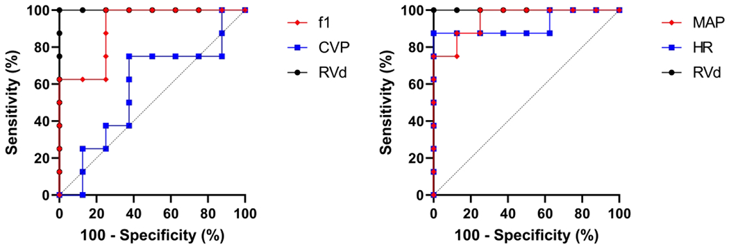
Peripheral intravenous analysis (PIVA) has been shown to be more sensitive than central venous pressure (CVP) for detecting hemorrhage and volume overload. We hypothesized that PIVA is superior to CVP for detecting right ventricular (RV) failure in a rat model of respiratory arrest.
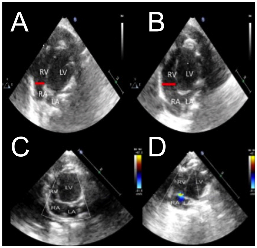
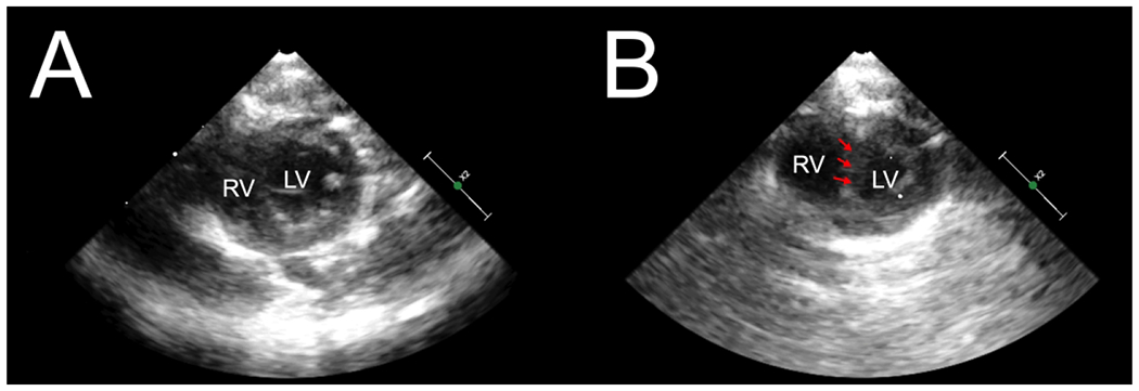
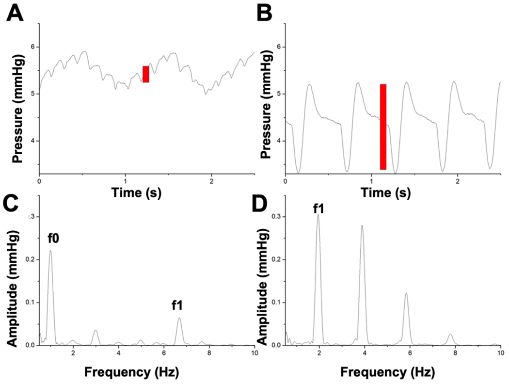
Eight Wistar rats were studied in accordance with the ARRIVE guidelines. CVP, mean arterial pressure (MAP), and PIVA were recorded. Respiratory arrest was achieved with IV Rocuronium. PIVA utilizes Fourier transform to quantify the amplitude of the peripheral venous waveform, expressed as the "f1 amplitude". RV diameter was measured with transthoracic echocardiography.
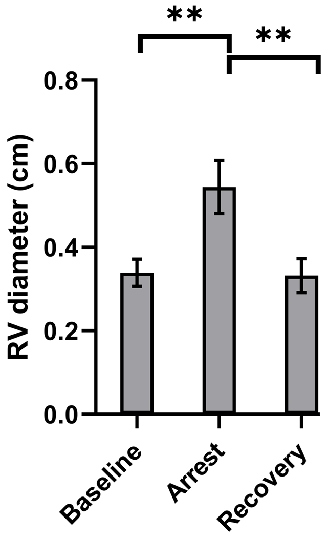
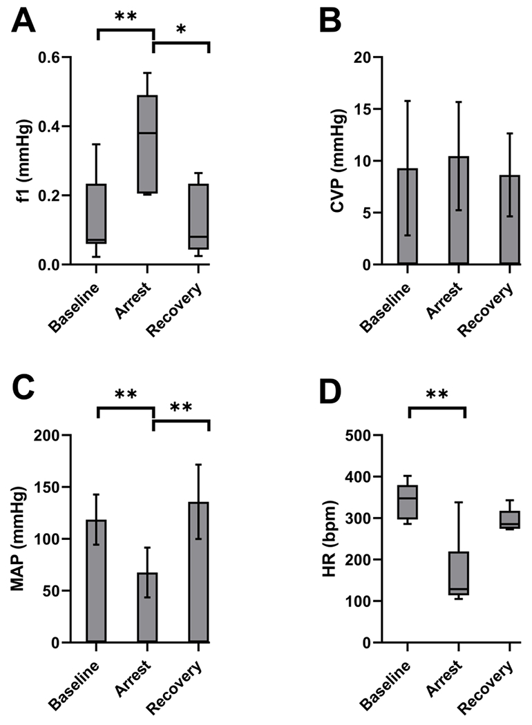
RV diameter increased from 0.34 to 0.54 cm during arrest, p = 0.001, and returned to 0.33 cm post arrest, p = 0.97. There was an increase in f1 amplitude from 0.07 to 0.38 mmHg, p = 0.01 and returned to 0.08 mmHg, p = 1.0. MAP decreased from 119 to 67 mmHg, p = 0.004 and returned to 136 mmHg, p = 0.50. There was no significant increase in CVP from 9.3 mmHg at baseline to 10.5 mmHg during respiratory arrest, p = 0.91, and recovery to 8.6 mmHg, p = 0.81.
This study highlights the utility of PIVA to detect RV failure in small-caliber vessels, comparable to peripheral veins in the human pediatric population.
Right ventricular failure remains a diagnostic challenge, particularly in pediatric patients with small vessel sizes limiting invasive intravascular monitor use. Intravenous analysis has shown promise in detecting hypovolemia and volume overload. Intravenous analysis successfully detects right ventricular failure in a rat respiratory arrest model. Intravenous analysis showed utility despite utilizing small peripheral venous access and therefore may be applicable to a pediatric population. Intravenous analysis may be helpful in differentiating various types of shock.
外周静脉分析(PIVA)在检测出血和容量超负荷方面比中心静脉压(CVP)更敏感。我们假设,在呼吸暂停的大鼠模型中,PIVA 比 CVP 更能检测到右心室(RV)衰竭。
根据 ARRIVE 指南,对 8 只 Wistar 大鼠进行了研究。记录 CVP、平均动脉压(MAP)和 PIVA。通过 IV 罗库溴铵实现呼吸暂停。PIVA 利用傅立叶变换来量化外周静脉波形的幅度,用“f1 幅度”表示。用经胸超声心动图测量 RV 直径。
RV 直径在呼吸暂停期间从 0.34 增加到 0.54cm,p=0.001,并在呼吸暂停后恢复到 0.33cm,p=0.97。f1 幅度从 0.07 增加到 0.38mmHg,p=0.01,恢复到 0.08mmHg,p=1.0。MAP 从 119 下降到 67mmHg,p=0.004,并恢复到 136mmHg,p=0.50。CVP 从基线时的 9.3mmHg 在呼吸暂停期间没有显著增加到 10.5mmHg,p=0.91,恢复到 8.6mmHg,p=0.81。
本研究强调了 PIVA 在检测小口径血管 RV 衰竭中的作用,与儿科人群中的外周静脉相当。
RV 衰竭仍然是一个诊断挑战,特别是在血管尺寸较小限制了侵入性血管内监测器使用的儿科患者中。静脉内分析在检测低血容量和容量超负荷方面显示出了希望。静脉内分析成功地在呼吸暂停的大鼠模型中检测到 RV 衰竭。尽管使用了小的外周静脉通路,静脉内分析仍具有实用性,因此可能适用于儿科人群。静脉内分析可能有助于区分各种类型的休克。