Lhuaire Martin, Wavreille Guillaume, Hivelin Mikael, Aumar Aurélien, Hunsinger Vincent, Derder Mohamed, Lellouch Alexandre G, Abrahams Peter, Lantieri Laurent, Fontaine Christian
Department of Plastic, Reconstructive and Aesthetic Surgery, Hôpital Européen Georges Pompidou, Assistance Publique des Hôpitaux de Paris, Université de Paris, Paris, France.
Institute of Anatomy and Organogenesis, Faculté de Médecine Henri Warembourg, Université de Lille, Lille, France.
JPRAS Open. 2022 May 14;33:171-183. doi: 10.1016/j.jpra.2022.04.008. eCollection 2022 Sep.
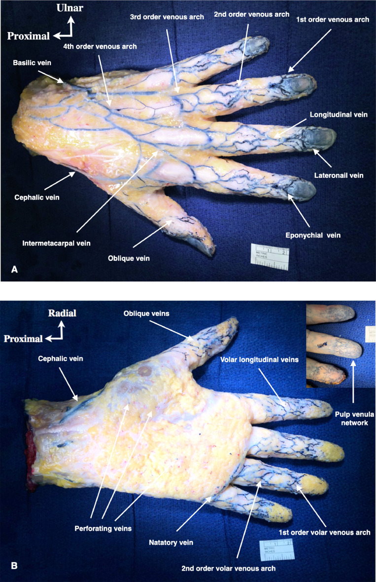
Venous anatomy of the digits and the hand is poorly reported in the literature compared to arterial anatomy. While knowledge of the venous anatomy is crucial to ensure safe skin incisions, skin flap design, or blood return restoration for digital replantations, data in anatomical and clinical textbooks are rather limited. The purpose of this anatomical study was to describe the venous anatomy of the digits and the hand.
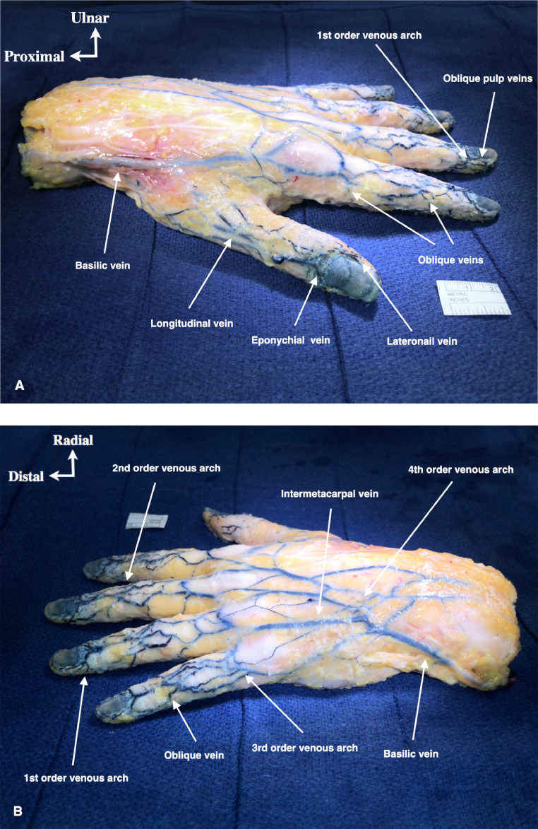
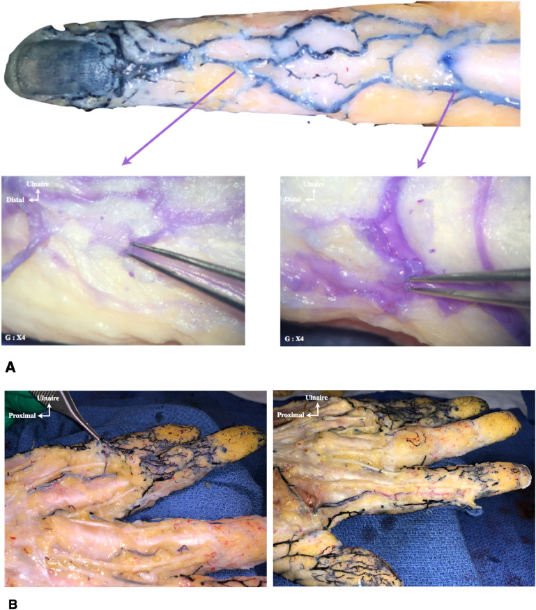
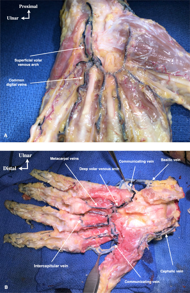
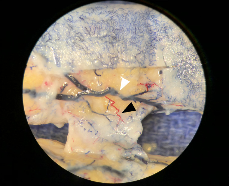
Our series reports descriptive results from 10 non-embalmed hand dissections from 5 different corpses. Hands were previously co-injected by arteries followed by veins with a different colored latex before being dissected under optical magnification (x4). Each anatomical specimen was photographed before being analyzed.
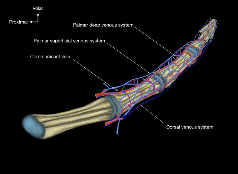
Each injection revealed both arterial and venous vascular systems. Latex injections were a useful technique to show the dorsal, volar superficial, and deep venous system. There was a constant and reliable topographic vascular anatomy of the superficial venous system of the digits and hand. However, we could not observe a high density of dorsal superficial venous valves as previously reported.
The knowledge of the arrangement of the venous system of the digits and the hand should help the surgeon when performing surgical procedures in the hand. The surgeon should take into consideration this venous anatomy when performing skin incisions, skin flaps, or replantation procedures which would preserve the normal venous physiology as much as possible.
与动脉解剖相比,文献中关于手指和手部静脉解剖的报道较少。虽然了解静脉解剖对于确保安全的皮肤切口、皮瓣设计或断指再植的血液回流恢复至关重要,但解剖学和临床教科书中的数据相当有限。本解剖学研究的目的是描述手指和手部的静脉解剖。
我们的系列报告了来自5具不同尸体的10次未防腐手部解剖的描述性结果。在光学放大(4倍)下进行解剖之前,先通过动脉然后用不同颜色的乳胶通过静脉对手部进行联合注射。每个解剖标本在分析之前都进行了拍照。
每次注射都显示了动脉和静脉血管系统。乳胶注射是显示手背、掌侧浅静脉和深静脉系统的有用技术。手指和手部浅静脉系统存在恒定且可靠的局部血管解剖结构。然而,我们没有观察到如先前报道的高密度手背浅静脉瓣膜。
了解手指和手部静脉系统的排列在外科医生进行手部手术时应有所帮助。外科医生在进行皮肤切口、皮瓣或再植手术时应考虑这种静脉解剖结构,尽可能保留正常的静脉生理功能。