Department of Radiology, Center for Advanced Imaging Innovation and Research (CAI2R), New York University Grossman School of Medicine, 660 First Ave, New York, NY, USA.
Medical Physics, Memorial Sloan Kettering Cancer Center, New York, NY, USA.
Sci Rep. 2022 Sep 2;12(1):15010. doi: 10.1038/s41598-022-19282-6.
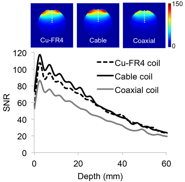
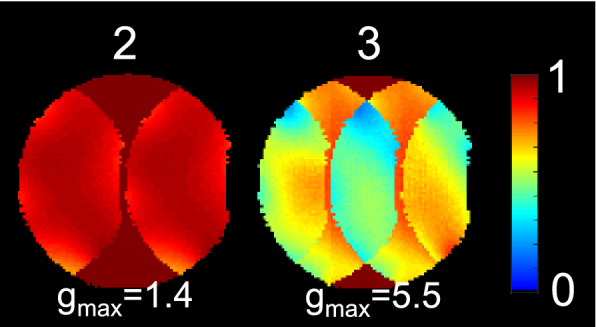
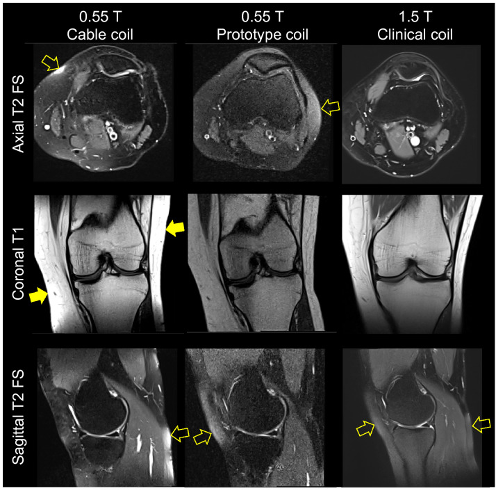
Flexible radiofrequency coils for magnetic resonance imaging (MRI) have garnered attention in research and industrial communities because they provide improved accessibility and performance and can accommodate a range of anatomic postures. Most recent flexible coil developments involve customized conductors or substrate materials and/or target applications at 3 T or above. In contrast, we set out to design a flexible coil based on an off-the-shelf conductor that is suitable for operation at 0.55 T (23.55 MHz). Signal-to-noise ratio (SNR) degradation can occur in such an environment because the resistance of the coil conductor can be significant with respect to the sample. We found that resonating a commercially available RG-223 coaxial cable shield with a lumped capacitor while the inner conductor remained electrically floating gave rise to a highly effective "cable coil." A 10-cm diameter cable coil was flexible enough to wrap around the knee, an application that can benefit from flexible coils, and had similar conductor loss and SNR as a standard-of-reference rigid copper coil. A two-channel cable coil array also provided good SNR robustness against geometric variability, outperforming a two-channel coaxial coil array by 26 and 16% when the elements were overlapped by 20-40% or gapped by 30-50%, respectively. A 6-channel cable coil array was constructed for 0.55 T knee imaging. Incidental cartilage and bone pathologies were clearly delineated in T1- and T2-weighted turbo spin echo images acquired in 3-4 min with the proposed coil, suggesting that clinical quality knee imaging is feasible in an acceptable examination timeframe. Correcting for T1, the SNR measured with the cable coil was approximately threefold lower than that measured with a 1.5 T state-of-the-art 18-channel coil, which is expected given the threefold difference in main magnetic field strength. This result suggests that the 0.55 T cable coil conductor loss does not deleteriously impact SNR, which might be anticipated at low field.
用于磁共振成像 (MRI) 的柔性射频线圈在研究和工业界引起了关注,因为它们提供了更好的可及性和性能,并可以适应各种解剖体位。最近的柔性线圈发展涉及定制导体或基板材料和/或在 3T 或更高的目标应用。相比之下,我们着手设计一种基于现成导体的柔性线圈,该导体适用于 0.55T(23.55MHz)操作。由于线圈导体的电阻相对于样品可能很大,因此可能会出现信噪比 (SNR) 下降。我们发现,在内部导体保持电浮的情况下,与集中电容器共振商用 RG-223 同轴电缆屏蔽会产生一种非常有效的“电缆线圈”。一个 10 厘米直径的电缆线圈足够灵活,可以缠绕在膝盖周围,这是一种可以从柔性线圈中受益的应用,并且具有与标准参考刚性铜线圈相似的导体损耗和 SNR。一个双通道电缆线圈阵列也提供了良好的 SNR 稳健性,能够抵抗几何变化,当元件重叠 20-40%或间隔 30-50%时,与双通道同轴线圈阵列相比,分别提高了 26%和 16%。构建了一个用于 0.55T 膝盖成像的 6 通道电缆线圈阵列。在用所提出的线圈在 3-4 分钟内采集的 T1 和 T2 加权涡轮自旋回波图像中,可以清楚地描绘出偶然的软骨和骨病理学,这表明在可接受的检查时间内可以进行临床质量的膝盖成像。校正 T1 后,用电缆线圈测量的 SNR 比用 1.5T 最先进的 18 通道线圈测量的 SNR 大约低三倍,这是由于主磁场强度的三倍差异所致。这一结果表明,0.55T 电缆线圈的导体损耗不会对 SNR 产生不利影响,这在低场中可能是预期的。