Computational Imaging Research Lab, Department of Biomedical Imaging and Image-guided Therapy, Medical University of Vienna, Spitalgasse 23, 1090, Vienna, Austria.
Department of Biomedical Imaging and Image-guided Therapy, Medical University of Vienna, Währinger Gürtel 18-20, 1090, Vienna, Austria.
Eur Radiol. 2023 Feb;33(2):925-935. doi: 10.1007/s00330-022-09101-x. Epub 2022 Sep 6.
To identify and evaluate predictive lung imaging markers and their pathways of change during progression of idiopathic pulmonary fibrosis (IPF) from sequential data of an IPF cohort. To test if these imaging markers predict outcome.
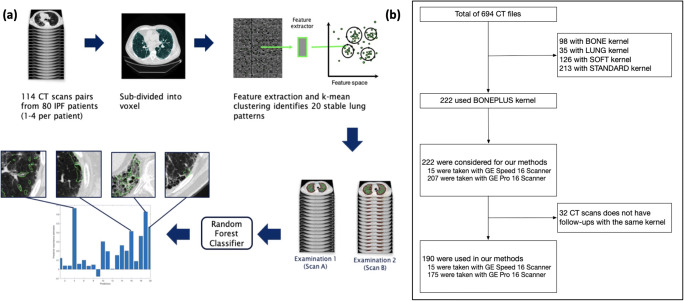
We studied radiological disease progression in 76 patients with IPF, including overall 190 computed tomography (CT) examinations of the chest. An algorithm identified candidates for imaging patterns marking progression by computationally clustering visual CT features. A classification algorithm selected clusters associated with radiological disease progression by testing their value for recognizing the temporal sequence of examinations. This resulted in radiological disease progression signatures, and pathways of lung tissue change accompanying progression observed across the cohort. Finally, we tested if the dynamics of marker patterns predict outcome, and performed an external validation study on a cohort from a different center.
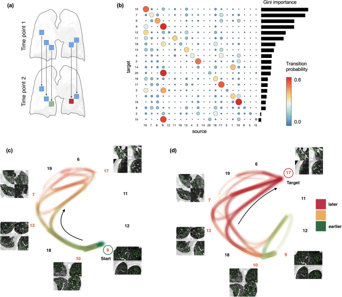
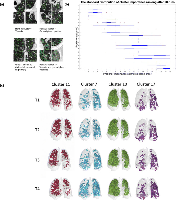
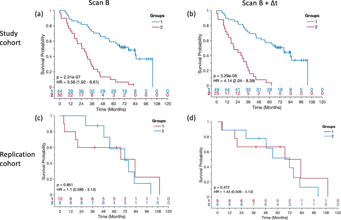
Progression marker patterns were identified and exhibited high stability in a repeatability experiment with 20 random sub-cohorts of the overall cohort. The 4 top-ranked progression markers were consistently selected as most informative for progression across all random sub-cohorts. After spatial image registration, local tracking of lung pattern transitions revealed a network of tissue transition pathways from healthy to a sequence of disease tissues. The progression markers were predictive for outcome, and the model achieved comparable results on a replication cohort.
Unsupervised learning can identify radiological disease progression markers that predict outcome. Local tracking of pattern transitions reveals pathways of radiological disease progression from healthy lung tissue through a sequence of diseased tissue types.
• Unsupervised learning can identify radiological disease progression markers that predict outcome in patients with idiopathic pulmonary fibrosis. • Local tracking of pattern transitions reveals pathways of radiological disease progression from healthy lung tissue through a sequence of diseased tissue types. • The progression markers achieved comparable results on a replication cohort.
从特发性肺纤维化(IPF)队列的连续数据中识别和评估预测性肺部成像标志物及其在纤维化进展过程中的变化途径,并检验这些成像标志物是否能预测结局。
我们研究了 76 例特发性肺纤维化患者的影像学疾病进展情况,共进行了 190 次胸部计算机断层扫描(CT)检查。一种算法通过对视觉 CT 特征进行计算聚类,确定了用于标记进展的成像模式候选者。分类算法通过检验其识别检查时间序列的能力,选择与影像学疾病进展相关的聚类。这就得到了整个队列中观察到的影像学疾病进展特征和伴随进展的肺组织变化途径。最后,我们检验了标志物模式的动态是否能预测结局,并在来自不同中心的队列上进行了外部验证研究。
在对整个队列的 20 个随机子队列进行的重复性实验中,确定了进展标志物模式,并表现出较高的稳定性。在所有随机子队列中,排名前 4 的进展标志物始终被选为最能反映进展的信息标志物。经过空间图像配准,对肺模式转变的局部跟踪揭示了从健康到一系列疾病组织的组织转变途径网络。进展标志物对结局具有预测作用,该模型在复制队列中也取得了类似的结果。
无监督学习可以识别预测特发性肺纤维化患者结局的影像学疾病进展标志物。对模式转变的局部跟踪揭示了从健康肺组织到一系列疾病组织类型的影像学疾病进展途径。
• 无监督学习可以识别预测特发性肺纤维化患者结局的影像学疾病进展标志物。• 对模式转变的局部跟踪揭示了从健康肺组织到一系列疾病组织类型的影像学疾病进展途径。• 该进展标志物在复制队列中取得了类似的结果。