Basaia Silvia, Agosta Federica, Francia Alessandro, Cividini Camilla, Balestrino Roberta, Stojkovic Tanja, Stankovic Iva, Markovic Vladana, Sarasso Elisabetta, Gardoni Andrea, De Micco Rosita, Albano Luigi, Stefanova Elka, Kostic Vladimir S, Filippi Massimo
Neuroimaging Research Unit, Division of Neuroscience, IRCCS San Raffaele Scientific Institute, Milan, Italy.
Neurology Unit, IRCCS San Raffaele Scientific Institute, Milan, Italy.
NPJ Parkinsons Dis. 2022 Sep 6;8(1):113. doi: 10.1038/s41531-022-00377-w.
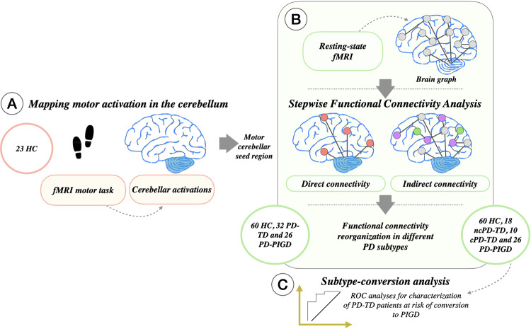
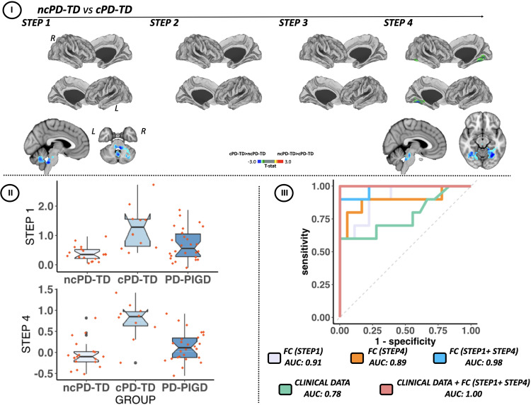
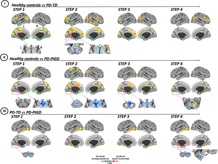
Parkinson's disease (PD) patients can be classified in tremor-dominant (TD) and postural-instability-and-gait-disorder (PIGD) motor subtypes. PIGD represents a more aggressive form of the disease that TD patients have a potentiality of converting into. This study investigated functional alterations within the cerebro-cerebellar system in PD-TD and PD-PIGD patients using stepwise functional connectivity (SFC) analysis and identified neuroimaging features that predict TD to PIGD conversion. Thirty-two PD-TD, 26 PD-PIGD patients and 60 healthy controls performed clinical/cognitive evaluations and resting-state functional MRI (fMRI). Four-year clinical follow-up data were available for 28 PD-TD patients, who were classified in 10 converters (cTD-PD) and 18 non-converters (ncTD-PD) to PIGD. The cerebellar seed-region was identified using a fMRI motor task. SFC analysis, characterizing regions that connect brain areas to the cerebellar seed at different levels of link-step distances, evaluated similar and divergent alterations in PD-TD and PD-PIGD. The discriminatory power of clinical data and/or SFC in distinguishing cPD-TD from ncPD-TD patients was assessed using ROC curve analysis. Compared to PD-TD, PD-PIGD patients showed decreased SFC in temporal lobe and occipital lobes and increased SFC in cerebellar cortex and ponto-medullary junction. Considering the subtype-conversion analysis, cPD-TD patients were characterized by increased SFC in temporal and occipital lobes and in cerebellum and ponto-medullary junction relative to ncPD-TD group. Combining clinical and SFC data, ROC curves provided the highest classification power to identify conversion to PIGD. These findings provide novel insights into the pathophysiology underlying different PD motor phenotypes and a potential tool for early characterization of PD-TD patients at risk of conversion to PIGD.
帕金森病(PD)患者可分为震颤为主型(TD)和姿势不稳与步态障碍型(PIGD)运动亚型。PIGD代表疾病的一种更具侵袭性的形式,TD患者有转化为该类型的可能性。本研究使用逐步功能连接(SFC)分析调查了PD-TD和PD-PIGD患者脑-小脑系统内的功能改变,并确定了预测TD向PIGD转化的神经影像学特征。32例PD-TD患者、26例PD-PIGD患者和60名健康对照者进行了临床/认知评估及静息态功能磁共振成像(fMRI)检查。28例PD-TD患者有4年的临床随访数据,这些患者被分为10例转化为PIGD的患者(cTD-PD)和18例未转化的患者(ncTD-PD)。使用fMRI运动任务确定小脑种子区域。SFC分析通过在不同链接步长距离水平上表征连接脑区与小脑种子区域的区域,评估了PD-TD和PD-PIGD中相似和不同的改变。使用ROC曲线分析评估临床数据和/或SFC区分cPD-TD和ncPD-TD患者的鉴别能力。与PD-TD相比,PD-PIGD患者颞叶和枕叶的SFC降低,小脑皮质和脑桥延髓交界处的SFC增加。考虑亚型转化分析,相对于ncPD-TD组,cPD-TD患者颞叶、枕叶、小脑和脑桥延髓交界处的SFC增加。结合临床和SFC数据,ROC曲线在识别向PIGD的转化方面具有最高的分类能力。这些发现为不同PD运动表型的病理生理学提供了新的见解,并为早期识别有转化为PIGD风险的PD-TD患者提供了潜在工具。