Faculty Campus Fryslân, University of Groningen, Leeuwarden, 8911 CE, The Netherlands.
Department of Surgery, Optical Molecular Imaging Groningen, University Medical Centre Groningen, Groningen, 9713 GZ, The Netherlands.
Surg Endosc. 2023 Feb;37(2):950-957. doi: 10.1007/s00464-022-09536-9. Epub 2022 Sep 6.
Ischemia at the site of an intestinal anastomosis is one of the most important risk factors for anastomotic leakage (AL). Consequently, adequate intestinal microperfusion is essential for optimal tissue oxygenation and anastomotic healing. As visual inspection of tissue viability does not guarantee an adequate objective evaluation of intestinal microperfusion, surgeons are in dire need of supportive tools to decrease anastomotic leakage after colorectal surgery.
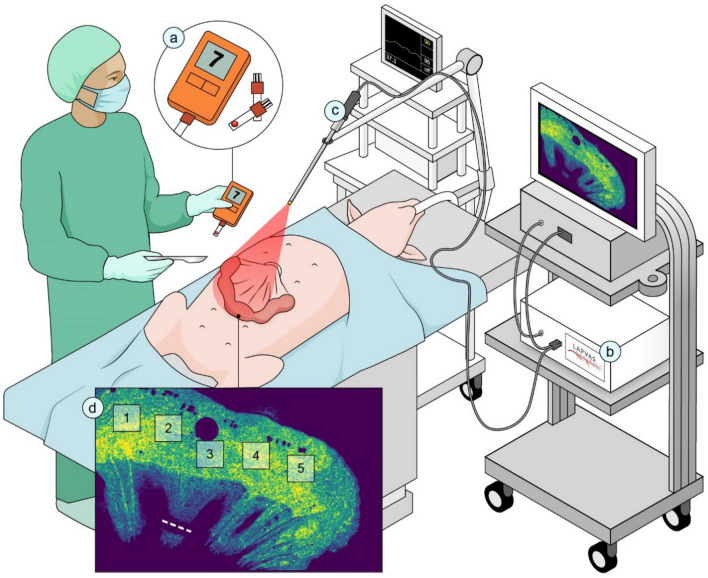
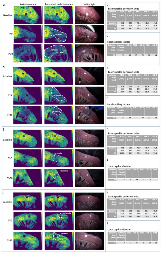
In this feasibility study, laparoscopic laser speckle contrast imaging (LSCI) was used to evaluate intestinal microperfusion in an experimental ischemic bowel loop model. Both large and small ischemic loops were created from the small intestine of a pig; each loop was divided into 5 regions of interest (ROI) with varying levels of ischemia. Speckle contrast and local capillary lactate (LCL) was measured in all ROIs.
Both real-time visualization of intestinal microperfusion and induced perfusion deficits was achieved in all bowel loops. As a result, the emergence of regions of intestinal ischemia could be predicted directly after iatrogenic perfusion limitation, whereas without LSCI signs of decreased intestinal viability could only be seen after 30 minutes. Additionally, a significant relation was found between LCL and LSCI.
In conclusion, LSCI can achieve real-time intraoperative visualization of intestinal microperfusion deficits, allowing for accurate prediction of long-term postoperative ischemic complications. With this revealing capacity, LSCI could potentially facilitate surgical decision-making when constructing intestinal anastomoses in order to mitigate ischemia-related complications such as AL.
肠吻合口缺血是吻合口漏(AL)的最重要危险因素之一。因此,充足的肠道微循环灌注对于最佳的组织氧合和吻合口愈合至关重要。由于组织活力的肉眼观察并不能保证对肠道微循环的充分客观评估,外科医生迫切需要支持工具来降低结直肠手术后的吻合口漏风险。
在这项可行性研究中,腹腔镜激光散斑对比成像(LSCI)用于评估实验性肠缺血环模型中的肠道微循环灌注。从小猪的小肠中创建大的和小的缺血环;每个环分为 5 个具有不同缺血程度的感兴趣区域(ROI)。在所有 ROI 中测量散斑对比度和局部毛细血管乳酸(LCL)。
所有肠环均实现了肠道微循环的实时可视化和诱导性灌注不足。因此,在医源性灌注受限后,可以直接预测肠缺血区域的出现,而没有 LSCI 则只能在 30 分钟后看到肠道活力下降的迹象。此外,还发现 LCL 与 LSCI 之间存在显著关系。
总之,LSCI 可以实现肠道微循环灌注不足的术中实时可视化,从而可以准确预测术后长期缺血性并发症。这种揭示能力可能有助于在构建肠吻合时进行手术决策,以减轻与缺血相关的并发症,如 AL。