• 文献检索
  • 文档翻译
  • 深度研究
  • 学术资讯
  • Suppr Zotero 插件Zotero 插件
  • 邀请有礼
  • 套餐&价格
  • 历史记录
应用&插件
Suppr Zotero 插件Zotero 插件浏览器插件Mac 客户端Windows 客户端微信小程序
定价
高级版会员购买积分包购买API积分包
服务
文献检索文档翻译深度研究API 文档MCP 服务
关于我们
关于 Suppr公司介绍联系我们用户协议隐私条款
关注我们

Suppr 超能文献

核心技术专利:CN118964589B侵权必究
粤ICP备2023148730 号-1Suppr @ 2026

文献检索

告别复杂PubMed语法,用中文像聊天一样搜索,搜遍4000万医学文献。AI智能推荐,让科研检索更轻松。

立即免费搜索

文件翻译

保留排版,准确专业,支持PDF/Word/PPT等文件格式,支持 12+语言互译。

免费翻译文档

深度研究

AI帮你快速写综述,25分钟生成高质量综述,智能提取关键信息,辅助科研写作。

立即免费体验

上皮性卵巢癌中存在大量的中心体缺陷。

A catalog of numerical centrosome defects in epithelial ovarian cancers.

机构信息

Biology of Centrosomes and Genetic Instability, Institut Curie, PSL Research University, CNRS UMR 144, Paris, France.

Department of Translational Research, Institut Curie, PSL University, Paris Cedex 05, France.

出版信息

EMBO Mol Med. 2022 Sep 7;14(9):e15670. doi: 10.15252/emmm.202215670.

DOI:10.15252/emmm.202215670
PMID:36069081
原文链接:https://pmc.ncbi.nlm.nih.gov/articles/PMC9449595/
Abstract

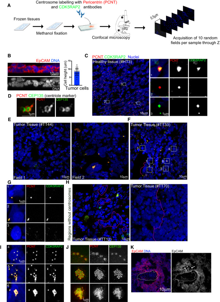
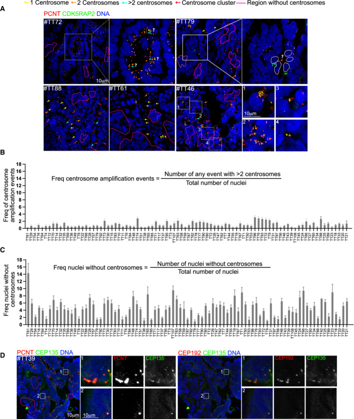
Centrosome amplification, the presence of more than two centrosomes in a cell is a common feature of most human cancer cell lines. However, little is known about centrosome numbers in human cancers and whether amplification or other numerical aberrations are frequently present. To address this question, we have analyzed a large cohort of primary human epithelial ovarian cancers (EOCs) from 100 patients. We found that rigorous quantitation of centrosome number in tumor samples was extremely challenging due to tumor heterogeneity and extensive tissue disorganization. Interestingly, even if centrosome clusters could be identified, the incidence of centrosome amplification was not comparable to what has been described in cultured cancer cells. Surprisingly, centrosome loss events where a few or many nuclei were not associated with centrosomes were clearly noticed and overall more frequent than centrosome amplification. Our findings highlight the difficulty of characterizing centrosome numbers in human tumors, while revealing a novel paradigm of centrosome number defects in EOCs.

摘要

中心体扩增,即一个细胞中存在两个以上的中心体,是大多数人类癌细胞系的一个常见特征。然而,关于人类癌症中的中心体数量以及扩增或其他数值异常是否经常存在,人们知之甚少。为了解决这个问题,我们分析了来自 100 名患者的大量原发性人上皮性卵巢癌 (EOC) 肿瘤样本。我们发现,由于肿瘤异质性和组织广泛紊乱,对肿瘤样本中中心体数量进行严格定量是极具挑战性的。有趣的是,即使可以识别中心体簇,中心体扩增的发生率也无法与在培养的癌细胞中所描述的相媲美。令人惊讶的是,中心体丢失事件,即少数或许多细胞核与中心体没有关联,明显被注意到,并且比中心体扩增更为常见。我们的研究结果强调了在人类肿瘤中描述中心体数量的困难,同时揭示了 EOC 中中心体数量缺陷的新范例。

https://cdn.ncbi.nlm.nih.gov/pmc/blobs/0e47/9449595/42a822208051/EMMM-14-e15670-g005.jpg
https://cdn.ncbi.nlm.nih.gov/pmc/blobs/0e47/9449595/6a8f1aa5dd78/EMMM-14-e15670-g004.jpg
https://cdn.ncbi.nlm.nih.gov/pmc/blobs/0e47/9449595/f0aaa4e41b68/EMMM-14-e15670-g001.jpg
https://cdn.ncbi.nlm.nih.gov/pmc/blobs/0e47/9449595/ecf160369f58/EMMM-14-e15670-g002.jpg
https://cdn.ncbi.nlm.nih.gov/pmc/blobs/0e47/9449595/42a822208051/EMMM-14-e15670-g005.jpg

相似文献

1
A catalog of numerical centrosome defects in epithelial ovarian cancers.上皮性卵巢癌中存在大量的中心体缺陷。
EMBO Mol Med. 2022 Sep 7;14(9):e15670. doi: 10.15252/emmm.202215670.
2
KIFCI, a novel putative prognostic biomarker for ovarian adenocarcinomas: delineating protein interaction networks and signaling circuitries.KIFCI,一种新型的卵巢腺癌潜在预后生物标志物:描绘蛋白质相互作用网络和信号通路。
J Ovarian Res. 2014 May 12;7:53. doi: 10.1186/1757-2215-7-53. eCollection 2014.
3
Centrosome abnormalities in ovarian cancer.卵巢癌中的中心体异常
Int J Cancer. 2005 Feb 20;113(5):746-51. doi: 10.1002/ijc.20633.
4
Centrosome amplification primes ovarian cancer cells for apoptosis and potentiates the response to chemotherapy.中心体扩增使卵巢癌细胞对细胞凋亡敏感,并增强对化疗的反应。
PLoS Biol. 2024 Sep 5;22(9):e3002759. doi: 10.1371/journal.pbio.3002759. eCollection 2024 Sep.
5
A centrosome clustering protein, KIFC1, predicts aggressive disease course in serous ovarian adenocarcinomas.一种中心体聚集蛋白KIFC1可预测浆液性卵巢腺癌的侵袭性病程。
J Ovarian Res. 2016 Mar 18;9:17. doi: 10.1186/s13048-016-0224-0.
6
The Impact of Centrosome Pathologies on Ovarian Cancer Development and Progression with a Focus on Centrosomes as Therapeutic Target.中心体病变对卵巢癌发生发展的影响及其作为治疗靶点的研究进展。
Adv Exp Med Biol. 2024;1452:37-64. doi: 10.1007/978-3-031-58311-7_3.
7
[The role of centrosomes in cells and their potential contribution to carcinogenesis].[中心体在细胞中的作用及其对致癌作用的潜在贡献]
Postepy Hig Med Dosw (Online). 2014 Jan 2;68:1050-68. doi: 10.5604/17322693.1119029.
8
Centrosome aberrations associated with cellular senescence and p53 localization at supernumerary centrosomes.与细胞衰老和多余中心体中 p53 定位相关的中心体异常。
Oxid Med Cell Longev. 2012;2012:217594. doi: 10.1155/2012/217594. Epub 2012 Oct 3.
9
A method of quantifying centrosomes at the single-cell level in human normal and cancer tissue.一种在人正常组织和肿瘤组织中单细胞水平定量中心体的方法。
Mol Biol Cell. 2019 Mar 21;30(7):811-819. doi: 10.1091/mbc.E18-10-0651. Epub 2019 Jan 30.
10
Intercellular centrosome number is correlated with the copy number of chromosomes in bladder cancer.膀胱癌细胞中的细胞间中心体数量与染色体拷贝数相关。
Cancer Genet Cytogenet. 2009 May;191(1):38-42. doi: 10.1016/j.cancergencyto.2009.01.012.

引用本文的文献

1
Screening a living biobank identifies cabazitaxel as a strategy to combat acquired taxol resistance in high-grade serous ovarian cancer.对一个活体生物样本库进行筛查后发现,卡巴他赛可作为一种策略来对抗高级别浆液性卵巢癌中获得性紫杉醇耐药。
Cell Rep Med. 2025 Jun 17;6(6):102160. doi: 10.1016/j.xcrm.2025.102160. Epub 2025 Jun 3.
2
Transient centrosome loss in cultured prostate epithelial cells induces chromosomal instability to produce an oncogenic genotype that correlates with poor clinical outcomes.培养的前列腺上皮细胞中短暂的中心体缺失会诱导染色体不稳定,产生与不良临床结果相关的致癌基因型。
bioRxiv. 2025 May 12:2025.05.10.653266. doi: 10.1101/2025.05.10.653266.
3

本文引用的文献

1
Centrosomes in disease: how the same music can sound so different?中心体与疾病:为何同曲异调?
Curr Opin Struct Biol. 2021 Feb;66:74-82. doi: 10.1016/j.sbi.2020.09.011. Epub 2020 Nov 10.
2
Differences in Mitotic Spindle Architecture in Mammalian Neural Stem Cells Influence Mitotic Accuracy during Brain Development.哺乳动物神经干细胞有丝分裂纺锤体结构的差异影响大脑发育过程中的有丝分裂准确性。
Curr Biol. 2019 Sep 23;29(18):2993-3005.e9. doi: 10.1016/j.cub.2019.07.061. Epub 2019 Sep 5.
3
Centrosome loss results in an unstable genome and malignant prostate tumors.
M phase-specific generation of supernumerary centrioles in cancer cells.
癌细胞中M期特异性产生的多余中心粒。
Mol Biol Cell. 2025 Jun 1;36(6):ar65. doi: 10.1091/mbc.E24-08-0386. Epub 2025 Apr 23.
4
Amplified centrosomes-more than just a threat.扩增的中心体——不仅仅是威胁。
EMBO Rep. 2024 Oct;25(10):4153-4167. doi: 10.1038/s44319-024-00260-0. Epub 2024 Sep 16.
5
Centrosome amplification primes ovarian cancer cells for apoptosis and potentiates the response to chemotherapy.中心体扩增使卵巢癌细胞对细胞凋亡敏感,并增强对化疗的反应。
PLoS Biol. 2024 Sep 5;22(9):e3002759. doi: 10.1371/journal.pbio.3002759. eCollection 2024 Sep.
6
Spindle assembly checkpoint-dependent mitotic delay is required for cell division in absence of centrosomes.纺锤体组装检验点依赖性有丝分裂延迟对于无中心体的细胞分裂是必需的。
Elife. 2024 Aug 2;12:RP84875. doi: 10.7554/eLife.84875.
7
Four centrosome-related genes to predict the prognosis and drug sensitivity of patients with colon cancer.四个与中心体相关的基因用于预测结肠癌患者的预后和药物敏感性。
World J Gastrointest Oncol. 2024 May 15;16(5):1908-1924. doi: 10.4251/wjgo.v16.i5.1908.
8
Cellular senescence in cancer: molecular mechanisms and therapeutic targets.癌症中的细胞衰老:分子机制与治疗靶点
MedComm (2020). 2024 Apr 24;5(5):e542. doi: 10.1002/mco2.542. eCollection 2024 May.
9
Molecular landscape and functional characterization of centrosome amplification in ovarian cancer.卵巢癌中中心体扩增的分子特征与功能研究
Nat Commun. 2023 Oct 16;14(1):6505. doi: 10.1038/s41467-023-41840-3.
10
The fate of extra centrosomes in newly formed tetraploid cells: should I stay, or should I go?新形成的四倍体细胞中额外中心体的命运:我该留下,还是离开?
Front Cell Dev Biol. 2023 Jul 27;11:1210983. doi: 10.3389/fcell.2023.1210983. eCollection 2023.
中心体缺失导致基因组不稳定和恶性前列腺肿瘤。
Oncogene. 2020 Jan;39(2):399-413. doi: 10.1038/s41388-019-0995-z. Epub 2019 Sep 2.
4
Plk4 Regulates Centriole Asymmetry and Spindle Orientation in Neural Stem Cells.Plk4 在神经干细胞中调节中心体不对称和纺锤体定向。
Dev Cell. 2019 Jul 1;50(1):11-24.e10. doi: 10.1016/j.devcel.2019.04.036. Epub 2019 May 23.
5
Oxidative Stress in Cells with Extra Centrosomes Drives Non-Cell-Autonomous Invasion.具有额外中心体的细胞中的氧化应激驱动非细胞自主侵袭。
Dev Cell. 2018 Nov 19;47(4):409-424.e9. doi: 10.1016/j.devcel.2018.10.026.
6
Molecular Mechanisms Regulating Organ-Specific Metastases in Epithelial Ovarian Carcinoma.调控上皮性卵巢癌器官特异性转移的分子机制
Cancers (Basel). 2018 Nov 15;10(11):444. doi: 10.3390/cancers10110444.
7
Over-elongation of centrioles in cancer promotes centriole amplification and chromosome missegregation.中心体在癌症中的过度延长促进了中心体扩增和染色体错误分离。
Nat Commun. 2018 Mar 28;9(1):1258. doi: 10.1038/s41467-018-03641-x.
8
Structural centrosome aberrations promote non-cell-autonomous invasiveness.结构中心体异常促进非细胞自主侵袭。
EMBO J. 2018 May 2;37(9). doi: 10.15252/embj.201798576. Epub 2018 Mar 22.
9
Author Correction: Cancer-associated fibroblasts induce metalloprotease-independent cancer cell invasion of the basement membrane.作者更正:癌症相关成纤维细胞诱导癌细胞不依赖金属蛋白酶侵袭基底膜。
Nat Commun. 2018 Mar 7;9(1):1036. doi: 10.1038/s41467-018-03304-x.
10
Olaparib tablets as maintenance therapy in patients with platinum-sensitive, relapsed ovarian cancer and a BRCA1/2 mutation (SOLO2/ENGOT-Ov21): a double-blind, randomised, placebo-controlled, phase 3 trial.奥拉帕利片作为 BRCA1/2 突变的铂敏感复发性卵巢癌患者的维持治疗(SOLO2/ENGOT-Ov21):一项双盲、随机、安慰剂对照、III 期临床试验。
Lancet Oncol. 2017 Sep;18(9):1274-1284. doi: 10.1016/S1470-2045(17)30469-2. Epub 2017 Jul 25.