Jiangxi Provincial Key Laboratory for Ophthalmology, Jiangxi Clinical Research Center of Ophthalmic Disease, Affiliated Eye Hospital of Nanchang University, Nanchang 330006, China.
Queen Mary School, Nanchang University, Nanchang 330006, China.
Cells. 2022 Aug 25;11(17):2645. doi: 10.3390/cells11172645.
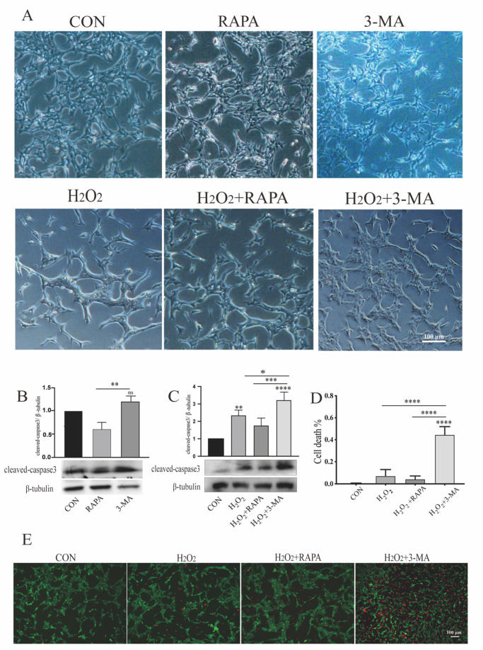
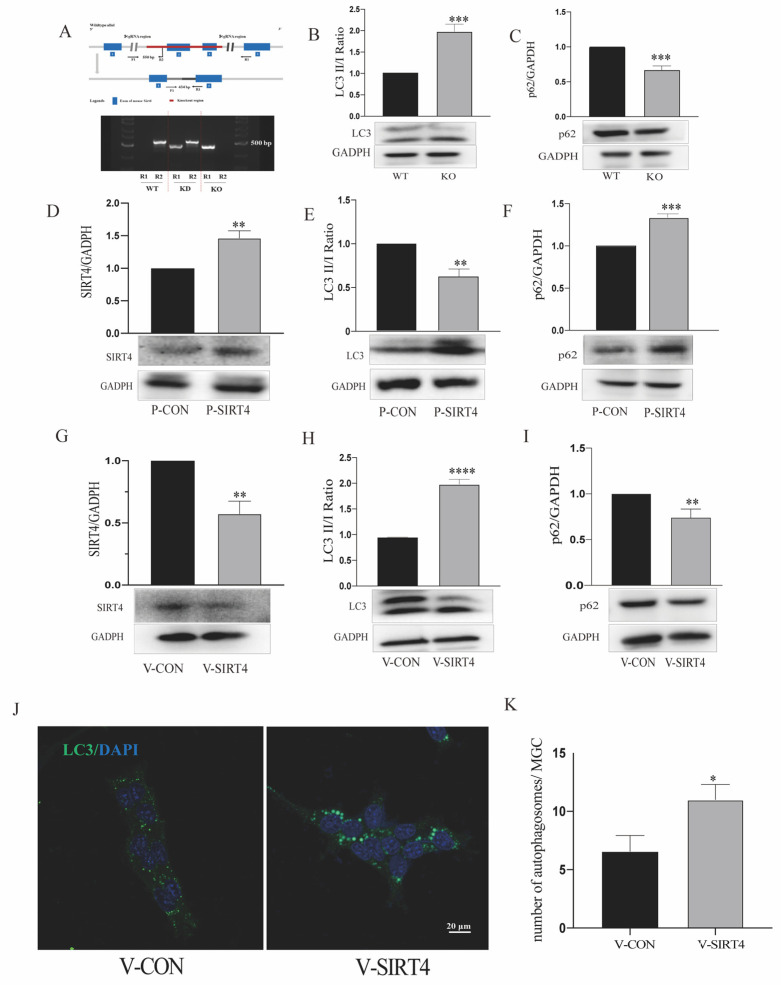
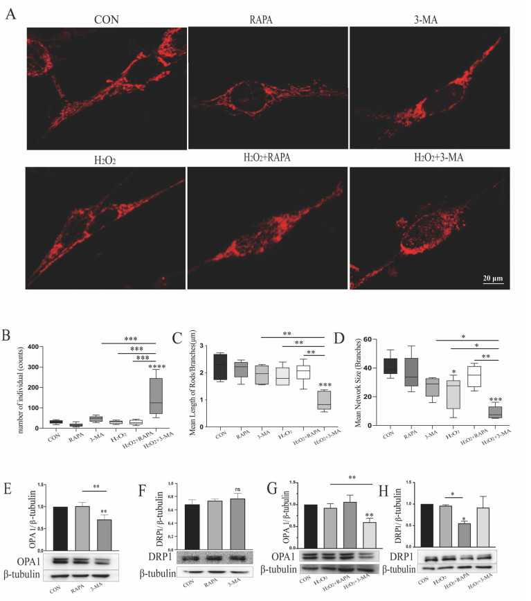
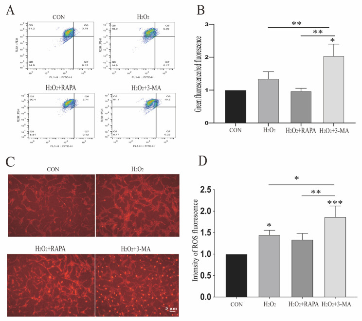
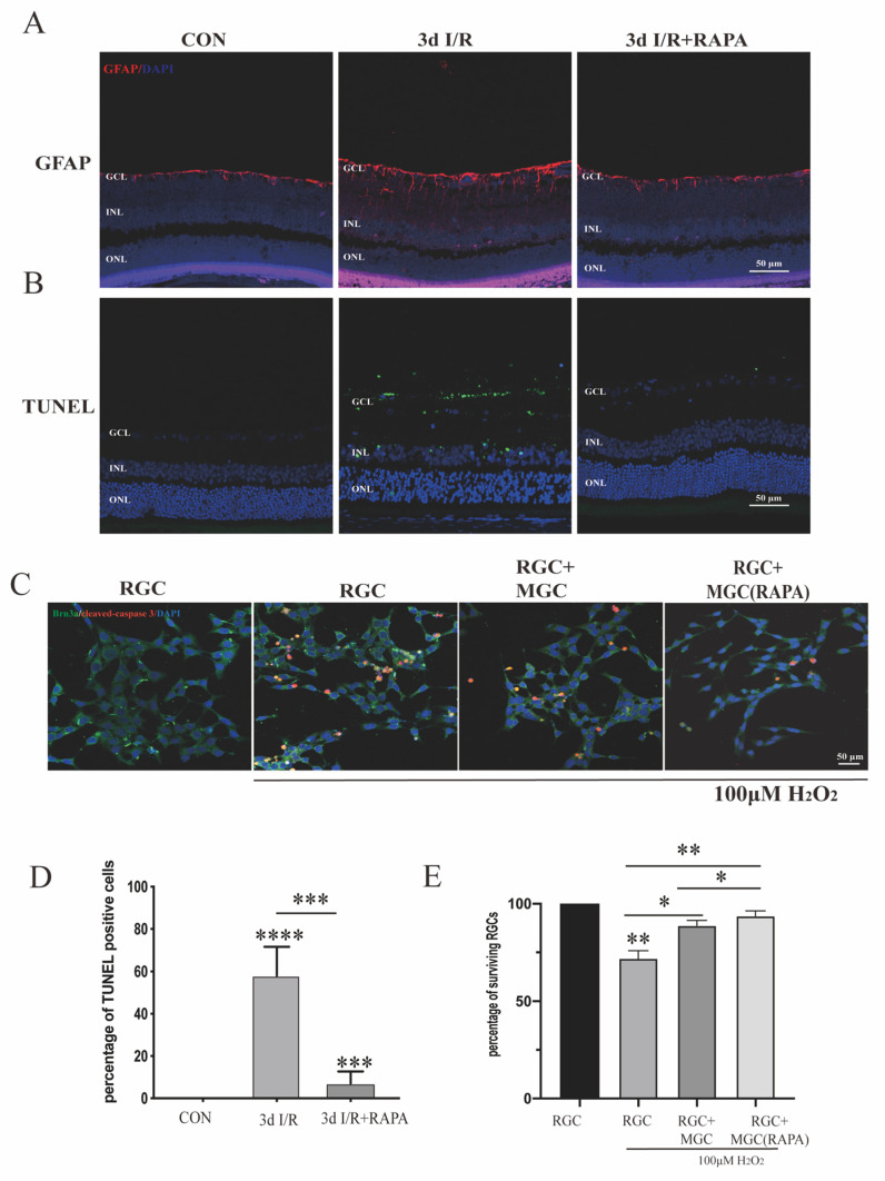
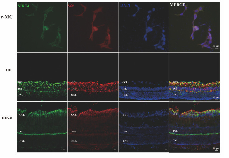
Müller glial cells (MGCs) are a group of glial cells in the retina that provide essential support to retinal neurons; however, the understanding of MGC apoptosis and autophagy remains limited. This study was aimed at investigating the role of autophagy in MGCs under normal and oxidative conditions, and identifying the underlying mechanisms. In addition, the sirtuin 4 (SIRT4)-mediated signaling pathway was observed to regulate the autophagic process in MGCs. To assess the effect of autophagy on MGC mitochondrial function and survival, we treated rMC-1 cells-rat-derived Müller glial cells-with rapamycin and 3-methyladenine (3-MA), and found that MGC death was not induced by such treatment, while autophagic dysfunction could increase MGC apoptosis under oxidative stress, as reflected by the expression level of cleaved caspase 3 and PI staining. In addition, the downregulation of autophagy by 3-MA could influence the morphology of the mitochondrial network structure, the mitochondrial membrane potential, and generation of reactive oxygen species (ROS) under oxidative stress. Moreover, SIRT4 depletion enhanced autophagosome formation, as verified by an increase in the LC3 II/I ratio and a decrease in the expression of SQSTM1/p62, and vice versa. The inhibition of AMPK phosphorylation by compound C could reverse these changes in LC3 II/I and SQSTM1/p62 caused by SIRT4 knockdown. Our research concludes that MGCs can endure autophagic dysfunction in the absence of oxidative stress, while the downregulation of autophagy can cause MGCs to become more sensitized to oxidative stress. Simultaneous exposure to oxidative stress and autophagic dysfunction in MGCs can result in a pronounced impairment of cell survival. Mechanically, SIRT4 depletion can activate the autophagic process in MGCs by regulating the AMPK-mTOR signaling pathway.
Muller 胶质细胞(MGC)是视网膜中的一组胶质细胞,为视网膜神经元提供必要的支持;然而,对 MGC 凋亡和自噬的理解仍然有限。本研究旨在探讨自噬在正常和氧化条件下对 MGC 的作用,并确定潜在的机制。此外,观察到 Sirtuin 4(SIRT4)介导的信号通路调节 MGC 中的自噬过程。为了评估自噬对 MGC 线粒体功能和存活的影响,我们用雷帕霉素和 3-甲基腺嘌呤(3-MA)处理 rMC-1 细胞-大鼠衍生的 Müller 胶质细胞-,发现这种处理不会诱导 MGC 死亡,而自噬功能障碍会增加 MGC 在氧化应激下的凋亡,这反映在 cleaved caspase 3 和 PI 染色的表达水平上。此外,3-MA 下调自噬会影响氧化应激下线粒体网络结构的形态、线粒体膜电位和活性氧(ROS)的产生。此外,SIRT4 耗竭通过增加 LC3 II/I 比值和降低 SQSTM1/p62 的表达来增强自噬体的形成,反之亦然。用化合物 C 抑制 AMPK 磷酸化可以逆转 SIRT4 敲低引起的 LC3 II/I 和 SQSTM1/p62 的这些变化。我们的研究得出结论,MGC 在没有氧化应激的情况下可以忍受自噬功能障碍,而下调自噬会使 MGC 对氧化应激更加敏感。同时暴露于 MGC 中的氧化应激和自噬功能障碍会导致细胞存活明显受损。在机制上,SIRT4 耗竭可以通过调节 AMPK-mTOR 信号通路激活 MGC 中的自噬过程。