Nittala Muneeswar G, Corvi Federico, Maram Jyotsna, Velaga Swetha B, Haines Jonathan, Pericak-Vance Margaret A, Stambolian Dwight, Sadda SriniVas R
Doheny Image Reading Center, Doheny Eye Institute, Los Angeles, CA 90033, USA.
Department of Epidemiology & Biostatistics, Case Western Reserve University, Cleveland, OH 44106, USA.
J Clin Med. 2022 Aug 30;11(17):5110. doi: 10.3390/jcm11175110.
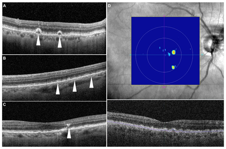
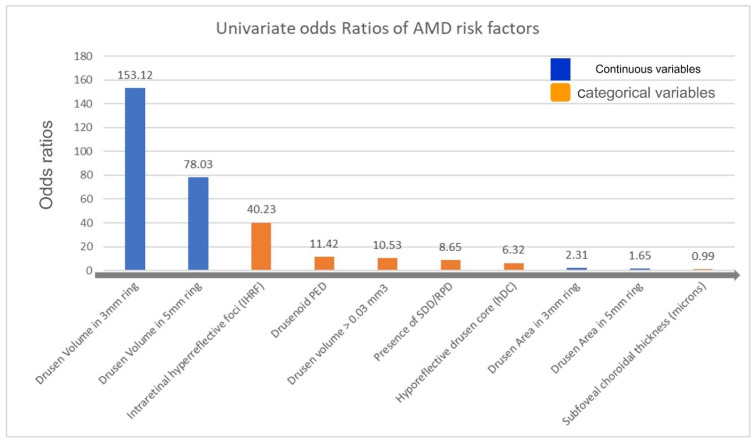
Objective: To evaluate the optical coherence tomography (OCT)-based risk factors for progression to late age-related macular degeneration (AMD) in a population-based study of elderly Amish. Methods: A total of 1332 eyes of 666 consecutive subjects who completed a 2-year follow-up visit were included in this multicenter, prospective, longitudinal, observational study. Imaging features were correlated with 2-year incidence of late AMD development. Odds ratios for imaging features were estimated from logistic regression. Baseline OCT images were reviewed for the presence of drusen volume ≥0.03 mm3 in the central 3 mm ring, intraretinal hyperreflective foci (IHRF), hyporeflective drusen cores (hDC), subretinal drusenoid deposits (SDD), and drusenoid pigment epithelium detachment (PED). Subfoveal choroidal thickness, drusen area, and drusen volume within 3 and 5 mm circles centered on the fovea were also assessed. Results: Twenty-one (1.5%) of 1332 eyes progressed to late AMD by 2 years. The mean age of the study subjects was 65 ± 10.17 (±SD) years and 410 subjects were female. Univariate logistic regression showed that drusen area and volume in both 3 mm and 5 mm circles, subfoveal choroidal thickness, drusen volume ≥ 0.03 mm3 in the 3 mm ring, SDD, IHRF, and hDC were all associated with an increased risk for development of late AMD. The multivariate regression model identified that drusen volume in the 3 mm ring (OR: 2.59, p = 0.049) and presence of IHRF (OR: 57.06, p < 0.001) remained as independent and significant risk factors for progression to late AMD. Conclusions: This population-based study confirms previous findings from clinic-based studies that high central drusen volume and IHRF are associated with an increased risk of progression to late AMD. These findings may be of value in risk-stratifying patients in clinical practice or identifying subjects for early intervention clinical trials.
在一项针对老年阿米什人的基于人群的研究中,评估基于光学相干断层扫描(OCT)的进展为晚期年龄相关性黄斑变性(AMD)的危险因素。方法:这项多中心、前瞻性、纵向观察性研究纳入了666名连续完成2年随访的受试者的1332只眼睛。将成像特征与晚期AMD发生的2年发病率相关联。成像特征的比值比通过逻辑回归估计。回顾基线OCT图像,观察中心3mm环内玻璃膜疣体积≥0.03mm³、视网膜内高反射灶(IHRF)、低反射玻璃膜疣核心(hDC)、视网膜下玻璃膜疣样沉积物(SDD)和玻璃膜疣样色素上皮脱离(PED)的情况。还评估了以黄斑中心凹为中心的3mm和5mm圆周内的黄斑下脉络膜厚度、玻璃膜疣面积和玻璃膜疣体积。结果:1332只眼睛中有21只(1.5%)在2年内进展为晚期AMD。研究对象的平均年龄为65±10.17(±标准差)岁,410名受试者为女性。单因素逻辑回归显示,3mm和5mm圆周内的玻璃膜疣面积和体积、黄斑下脉络膜厚度、3mm环内玻璃膜疣体积≥0.03mm³、SDD、IHRF和hDC均与晚期AMD发生风险增加相关。多变量回归模型确定,3mm环内的玻璃膜疣体积(比值比:2.59,p = 0.049)和IHRF的存在(比值比:57.06,p < 0.001)仍然是进展为晚期AMD的独立且显著的危险因素。结论:这项基于人群的研究证实了先前基于临床研究的结果,即高中心玻璃膜疣体积和IHRF与进展为晚期AMD的风险增加相关。这些发现可能在临床实践中对患者进行风险分层或识别早期干预临床试验的受试者方面具有价值。