• 文献检索
  • 文档翻译
  • 深度研究
  • 学术资讯
  • Suppr Zotero 插件Zotero 插件
  • 邀请有礼
  • 套餐&价格
  • 历史记录
应用&插件
Suppr Zotero 插件Zotero 插件浏览器插件Mac 客户端Windows 客户端微信小程序
定价
高级版会员购买积分包购买API积分包
服务
文献检索文档翻译深度研究API 文档MCP 服务
关于我们
关于 Suppr公司介绍联系我们用户协议隐私条款
关注我们

Suppr 超能文献

核心技术专利:CN118964589B侵权必究
粤ICP备2023148730 号-1Suppr @ 2026

文献检索

告别复杂PubMed语法,用中文像聊天一样搜索,搜遍4000万医学文献。AI智能推荐,让科研检索更轻松。

立即免费搜索

文件翻译

保留排版,准确专业,支持PDF/Word/PPT等文件格式,支持 12+语言互译。

免费翻译文档

深度研究

AI帮你快速写综述,25分钟生成高质量综述,智能提取关键信息,辅助科研写作。

立即免费体验

站立位 MRI 中冗余神经根的发生率在仰卧位时减少一半,在屈髋坐位时几乎降为零:一项回顾性的横断面队列研究。

The prevalence of redundant nerve roots in standing positional MRI decreases by half in supine and almost to zero in flexed seated position: a retrospective cross-sectional cohort study.

机构信息

Clinic for Spine Surgery, Schoen-Clinic Hamburg, Academic Hospital of the University Medical Center Eppendorf, Dehnhaide 120, 22081, Hamburg, Germany.

CRO - Dr. med. Kottmann GmbH & Co. KG, Hamm, Germany.

出版信息

Neuroradiology. 2022 Nov;64(11):2191-2201. doi: 10.1007/s00234-022-03047-z. Epub 2022 Sep 9.

DOI:10.1007/s00234-022-03047-z
PMID:36083504
原文链接:https://pmc.ncbi.nlm.nih.gov/articles/PMC9576640/
Abstract

PURPOSE

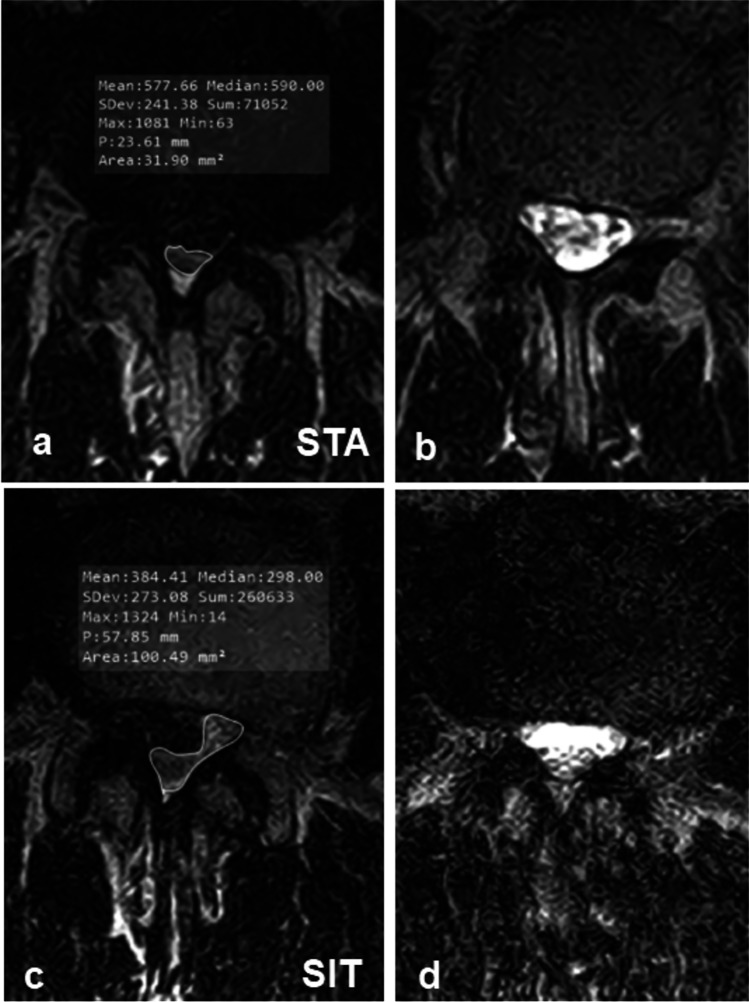
This retrospective cross-sectional cohort study investigated the influence of posture on lordosis (LL), length of the spinal canal (LSC), anteroposterior diameter (APD L1-L5), dural cross-sectional area (DCSA) of the lumbar spinal canal, and the prevalence of redundant nerve roots (RNR) using positional magnetic resonance imaging (MRI) (0.6 T).

METHODS

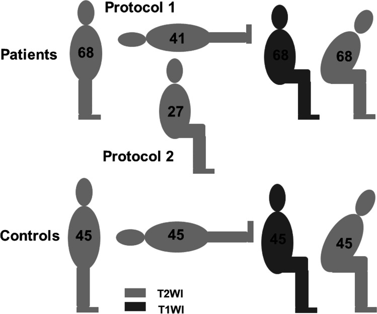
Sixty-eight patients with single-level degenerative central lumbar spinal stenosis (cLSS) presenting with RNR in the standing position (STA) were also investigated in supine (SUP) or neutral seated (SIT) and flexed seated (FLEX) positions. Additionally, 45 patients complaining of back pain and without MRI evidence of LSS were evaluated. Statistical significance was set at p < 0.05.

RESULTS

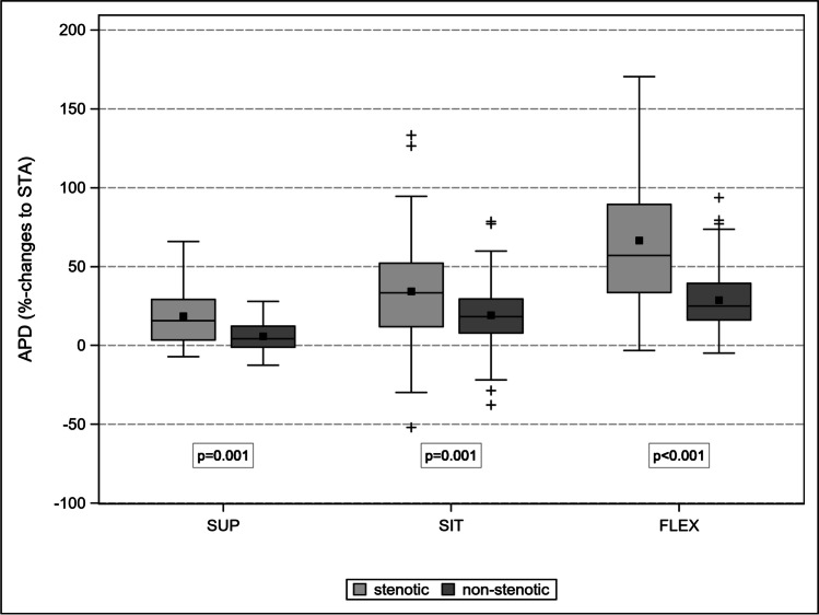
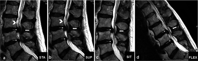
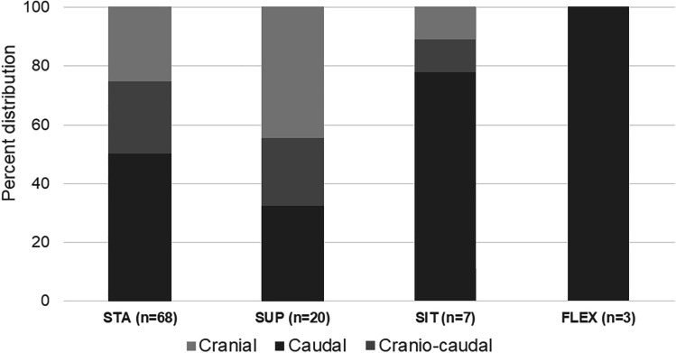
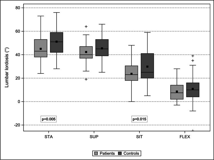
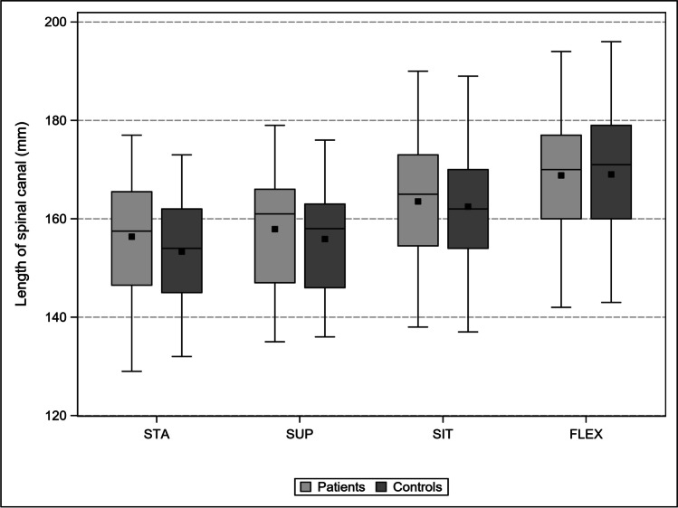
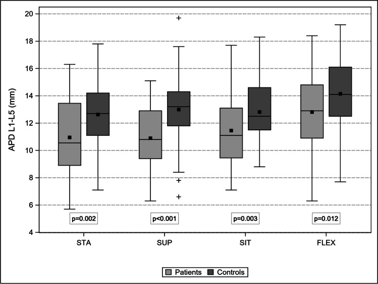
Controls (A) and patients with cLSS (B) were comparable in terms of mean age (p = 0.88) and sex (p = 0.22). The progressive transition from STA to FLEX led to a comparable decrease in LL (p = 0.97), an increase in LSC (p = 0.80), and an increase in APD L1-L5 (p = 0.78). The APD of the stenotic level increased disproportionally between the different postures, up to 67% in FLEX compared to 29% in adjacent non-stenotic levels (p < 0.001). Therefore, the prevalence of RNR decreased to 49, 26, and 4% in SUP, SIT, and FLEX, respectively.

CONCLUSION

The prevalence of RNR in standing position was underestimated by half in supine position. Body postures modified LL, LSC, and APD similarly in patients and controls. Stenotic levels compensated for insufficient intraspinal volume with a disproportionate enlargement when switching from the STA to FLEX.

摘要

目的

本回顾性横断面队列研究旨在通过体位磁共振成像(0.6T)研究体位对腰椎前凸(LL)、椎管长度(LSC)、前后径(L1-L5 的 APD)、椎管硬脊膜横截面积(DCSA)以及冗余神经根(RNR)的发生率的影响。

方法

本研究共纳入 68 例单节段退行性中央型腰椎管狭窄症(cLSS)患者,这些患者在站立位(STA)时存在 RNR,并在仰卧位(SUP)、中立位坐姿(SIT)和前屈位坐姿(FLEX)下进行检查。此外,还评估了 45 例腰痛但 MRI 未见 LSS 证据的患者。统计学意义设定为 p<0.05。

结果

cLSS 患者(B)与对照组(A)在平均年龄(p=0.88)和性别(p=0.22)方面具有可比性。STA 到 FLEX 的逐步过渡导致 LL 相似下降(p=0.97)、LSC 相似增加(p=0.80)以及 L1-L5 的 APD 相似增加(p=0.78)。在不同的体位下,狭窄水平的 APD 不成比例地增加,与相邻非狭窄水平相比,FLEX 时增加了 67%,而在 STA 时仅增加了 29%(p<0.001)。因此,SUP、SIT 和 FLEX 时 RNR 的发生率分别下降至 49%、26%和 4%。

结论

仰卧位时,站立位 RNR 的发生率被低估了一半。体位改变同样影响患者和对照组的 LL、LSC 和 APD。当从 STA 转换为 FLEX 时,狭窄水平通过不成比例的增大来补偿椎管内空间不足。

https://cdn.ncbi.nlm.nih.gov/pmc/blobs/e872/9576640/895bdf4b8321/234_2022_3047_Fig9_HTML.jpg
https://cdn.ncbi.nlm.nih.gov/pmc/blobs/e872/9576640/cfe706020800/234_2022_3047_Fig1_HTML.jpg
https://cdn.ncbi.nlm.nih.gov/pmc/blobs/e872/9576640/430a2338fc3a/234_2022_3047_Fig2_HTML.jpg
https://cdn.ncbi.nlm.nih.gov/pmc/blobs/e872/9576640/a19b029d91a5/234_2022_3047_Fig3_HTML.jpg
https://cdn.ncbi.nlm.nih.gov/pmc/blobs/e872/9576640/b70c658f3156/234_2022_3047_Fig4_HTML.jpg
https://cdn.ncbi.nlm.nih.gov/pmc/blobs/e872/9576640/0d6105683f8f/234_2022_3047_Fig5_HTML.jpg
https://cdn.ncbi.nlm.nih.gov/pmc/blobs/e872/9576640/5c8961e15a81/234_2022_3047_Fig6_HTML.jpg
https://cdn.ncbi.nlm.nih.gov/pmc/blobs/e872/9576640/b795be7af456/234_2022_3047_Fig7_HTML.jpg
https://cdn.ncbi.nlm.nih.gov/pmc/blobs/e872/9576640/ff8e4924c42e/234_2022_3047_Fig8_HTML.jpg
https://cdn.ncbi.nlm.nih.gov/pmc/blobs/e872/9576640/895bdf4b8321/234_2022_3047_Fig9_HTML.jpg

相似文献

1
The prevalence of redundant nerve roots in standing positional MRI decreases by half in supine and almost to zero in flexed seated position: a retrospective cross-sectional cohort study.站立位 MRI 中冗余神经根的发生率在仰卧位时减少一半,在屈髋坐位时几乎降为零:一项回顾性的横断面队列研究。
Neuroradiology. 2022 Nov;64(11):2191-2201. doi: 10.1007/s00234-022-03047-z. Epub 2022 Sep 9.
2
The prevalence of redundant nerve roots in patients with lumbar spinal stenosis is body position dependent: a retrospective observational study with repeated measures design in an upright MRI scanner.腰椎管狭窄症患者冗余神经根的患病率与体位有关:一项在直立 MRI 扫描仪中采用重复测量设计的回顾性观察研究。
Neuroradiology. 2020 Aug;62(8):979-985. doi: 10.1007/s00234-020-02423-x. Epub 2020 Apr 21.
3
Conventional Supine MRI With a Lumbar Pillow-An Alternative to Weight-bearing MRI for Diagnosing Spinal Stenosis?: A Cross-sectional Study.使用腰枕的传统仰卧位磁共振成像:诊断腰椎管狭窄症时负重磁共振成像的替代方法?一项横断面研究。
Spine (Phila Pa 1976). 2017 May 1;42(9):662-669. doi: 10.1097/BRS.0000000000001889.
4
Postural changes of the dural sac in the lumbar spines of asymptomatic individuals using positional stand-up magnetic resonance imaging.使用站立位磁共振成像观察无症状个体腰椎硬脊膜囊的体位变化。
Spine (Phila Pa 1976). 2007 Feb 15;32(4):E136-40. doi: 10.1097/01.brs.0000255202.94153.ca.
5
Patient demographics and MRI-based measurements predict redundant nerve roots in lumbar spinal stenosis: a retrospective database cohort comparison.患者人口统计学特征和基于磁共振成像的测量可预测腰椎管狭窄症中的神经根冗余:一项回顾性数据库队列比较研究
BMC Musculoskelet Disord. 2018 Dec 22;19(1):452. doi: 10.1186/s12891-018-2364-4.
6
Influence of posture on relationships between pelvic parameters and lumbar lordosis: Comparison of the standing, seated, and supine positions. A preliminary study.姿势对骨盆参数与腰椎前凸关系的影响:站立位、坐位和仰卧位的比较。初步研究。
Orthop Traumatol Surg Res. 2018 Sep;104(5):565-568. doi: 10.1016/j.otsr.2018.06.005. Epub 2018 Jul 31.
7
The effect of body position and axial load on spinal canal morphology: an MRI study of central spinal stenosis.体位和轴向负荷对椎管形态的影响:一项关于中央型椎管狭窄的MRI研究
Spine (Phila Pa 1976). 2008 Jan 1;33(1):61-7. doi: 10.1097/BRS.0b013e31815e395f.
8
Correlation Between Dural Sac Size in Dynamic Magnetic Resonance Imaging and Clinical Symptoms in Patients with Lumbar Spinal Stenosis.动态磁共振成像中硬脊膜囊大小与腰椎管狭窄症患者临床症状的相关性。
World Neurosurg. 2020 Feb;134:e866-e873. doi: 10.1016/j.wneu.2019.11.011. Epub 2019 Nov 9.
9
Lumbosacral spinal compression device with the use of a cushion back support in supine MRI.腰骶部脊柱压缩装置,在仰卧 MRI 中使用靠垫背部支撑。
Acta Radiol. 2021 Aug;62(8):1052-1062. doi: 10.1177/0284185120951963. Epub 2020 Aug 27.
10
Axial loading during supine MRI for improved assessment of lumbar spine: comparison with standing MRI.仰卧位 MRI 轴向加载改善腰椎评估:与站立位 MRI 的比较。
Acta Radiol. 2023 Jan;64(1):217-227. doi: 10.1177/02841851211068148. Epub 2021 Dec 23.

引用本文的文献

1
Can the supine extended position with legs straightened and a lumbar pillow complement the psoas-relaxed position for the diagnosis of lumbar spinal stenosis on MRI? A prospective cross-sectional study.双腿伸直的仰卧伸展位并使用腰垫,能否辅助腰大肌放松位用于磁共振成像(MRI)诊断腰椎管狭窄?一项前瞻性横断面研究。
Brain Spine. 2025 Aug 2;5:104377. doi: 10.1016/j.bas.2025.104377. eCollection 2025.
2
Lumbar spinal stenosis and surgical decompression affect sleep quality and position in patients. A prospective cross-sectional cohort study.腰椎管狭窄症及手术减压对患者睡眠质量和睡眠姿势的影响。一项前瞻性横断面队列研究。
Brain Spine. 2024 Mar 22;4:102785. doi: 10.1016/j.bas.2024.102785. eCollection 2024.
3

本文引用的文献

1
Reliability of reporting differences in degenerative MRI findings of the lumbar spine from the supine to the upright position.仰卧位到直立位腰椎退变 MRI 表现差异报告的可靠性。
Skeletal Radiol. 2022 Nov;51(11):2141-2154. doi: 10.1007/s00256-022-04060-2. Epub 2022 May 10.
2
Magnetic resonance imaging findings of redundant nerve roots of the cauda equina.马尾神经冗余神经根的磁共振成像表现
World J Radiol. 2021 Jan 28;13(1):29-39. doi: 10.4329/wjr.v13.i1.29.
3
The prevalence of redundant nerve roots in patients with lumbar spinal stenosis is body position dependent: a retrospective observational study with repeated measures design in an upright MRI scanner.
Epidural pressure measurement using a fiber-optic sensor (proof-of-principle in vivo animal trial).
使用光纤传感器进行硬膜外压力测量(体内动物试验原理验证)。
Animal Model Exp Med. 2024 Oct;7(5):769-776. doi: 10.1002/ame2.12469. Epub 2024 Jul 9.
4
MRI parameters predict central lumbar spinal stenosis combined with redundant nerve roots: a prospective MRI study.MRI参数预测中央型腰椎管狭窄合并神经根冗余:一项前瞻性MRI研究
Front Neurol. 2024 May 27;15:1385770. doi: 10.3389/fneur.2024.1385770. eCollection 2024.
腰椎管狭窄症患者冗余神经根的患病率与体位有关:一项在直立 MRI 扫描仪中采用重复测量设计的回顾性观察研究。
Neuroradiology. 2020 Aug;62(8):979-985. doi: 10.1007/s00234-020-02423-x. Epub 2020 Apr 21.
4
Redundant nerve roots in lumbar spinal stenosis: inter- and intra-rater reliability of an MRI-based classification.腰椎管狭窄症中冗余神经根:基于 MRI 的分类的组内和组间可靠性。
Neuroradiology. 2020 Feb;62(2):223-230. doi: 10.1007/s00234-019-02337-3. Epub 2019 Dec 14.
5
The clinical significance of redundant nerve roots of the cauda equina in lumbar spinal stenosis patients: A systematic literature review and meta-analysis.腰椎管狭窄症患者马尾神经冗余神经根的临床意义:一项系统文献综述与荟萃分析
Clin Neurol Neurosurg. 2018 Nov;174:40-47. doi: 10.1016/j.clineuro.2018.09.001. Epub 2018 Sep 4.
6
Dynamic MR in patients affected by neurogenical claudication: technique and results from a single-center experience.神经源性间歇性跛行患者的动态磁共振成像:单中心经验的技术与结果
Neuroradiology. 2016 Aug;58(8):765-70. doi: 10.1007/s00234-016-1697-7. Epub 2016 May 21.
7
Post-surgical functional recovery, lumbar lordosis, and range of motion associated with MR-detectable redundant nerve roots in lumbar spinal stenosis.腰椎管狭窄症中与磁共振成像(MR)可检测到的冗余神经根相关的术后功能恢复、腰椎前凸和活动范围。
Clin Neurol Neurosurg. 2016 Jan;140:79-84. doi: 10.1016/j.clineuro.2015.11.016. Epub 2015 Nov 24.
8
Intrathecal volume changes in lumbar spinal canal stenosis following extension and flexion: An experimental cadaver study.腰椎管狭窄症患者在伸展和屈曲后鞘内体积变化:一项尸体实验研究。
Technol Health Care. 2015;23(5):645-52. doi: 10.3233/THC-151011.
9
The Lumbar Spine as a Dynamic Structure Depicted in Upright MRI.直立位MRI所显示的作为动态结构的腰椎
Medicine (Baltimore). 2015 Aug;94(32):e1299. doi: 10.1097/MD.0000000000001299.
10
Redundant nerve roots of the cauda equina in lumbar spinal canal stenosis, an MR study on 500 cases.腰椎管狭窄症中马尾神经冗余神经根:500例的磁共振成像研究
Eur Spine J. 2015 Oct;24(10):2315-20. doi: 10.1007/s00586-015-4059-y. Epub 2015 Jun 14.