Vascular Biology Research Centre, Institute of Molecular and Clinical Sciences, St George's University of London, London, UK.
Mass Spectrometry Core Laboratory, Edinburgh Clinical Research Facility, Queen's Medical Research Institute, University of Edinburgh, Edinburgh, UK.
Br J Pharmacol. 2023 Jan;180(2):174-193. doi: 10.1111/bph.15947. Epub 2022 Oct 11.
Kcnq-encoded K 7 channels (termed K 7.1-5) regulate vascular smooth muscle cell (VSMC) contractility at rest and as targets of receptor-mediated responses. However, the current data are mostly derived from males. Considering the known effects of sex, the oestrous cycle and sex hormones on vascular reactivity, here we have characterised the molecular and functional properties of K 7 channels from renal and mesenteric arteries from female Wistar rats separated into di-oestrus and met-oestrus (F-D/M) and pro-oestrus and oestrus (F-P/E).
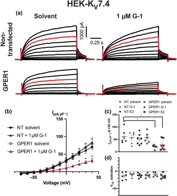
RT-qPCR, immunocytochemistry, proximity ligation assay and wire myography were performed in renal and mesenteric arteries. Circulating sex hormone concentrations were determined by liquid chromatography-tandem mass spectrometry. Whole-cell electrophysiology was undertaken on cells expressing K 7.4 channels in association with G-protein-coupled oestrogen receptor 1 (GPER1).
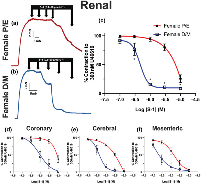
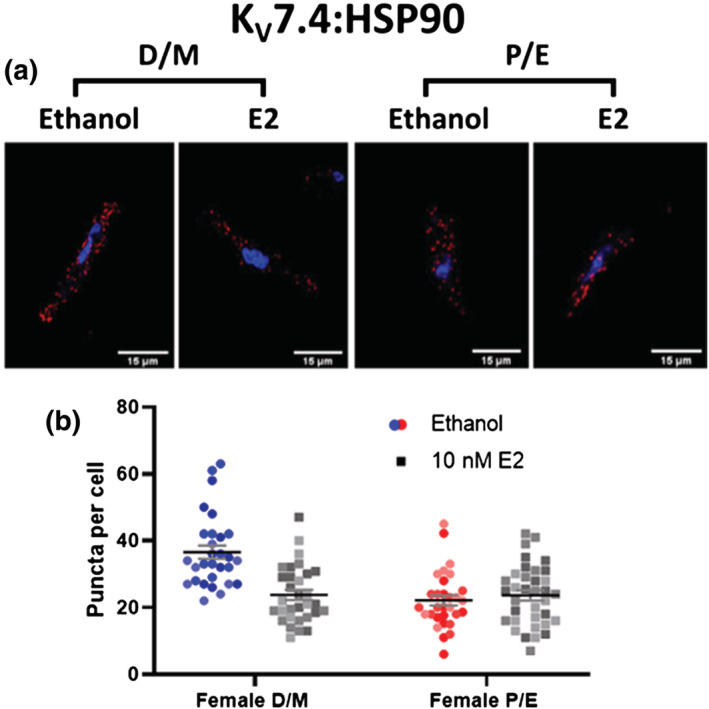
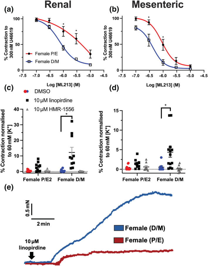
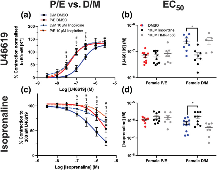
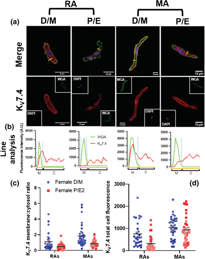
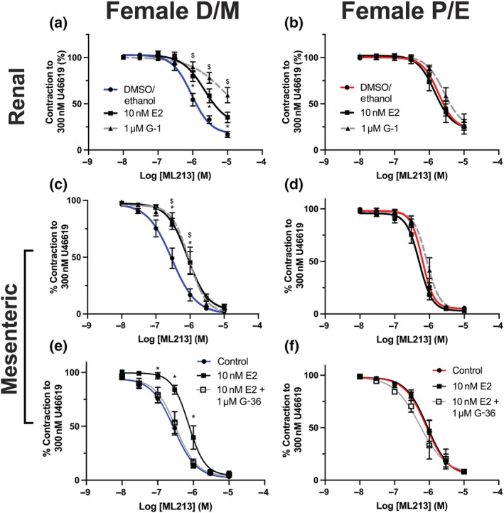
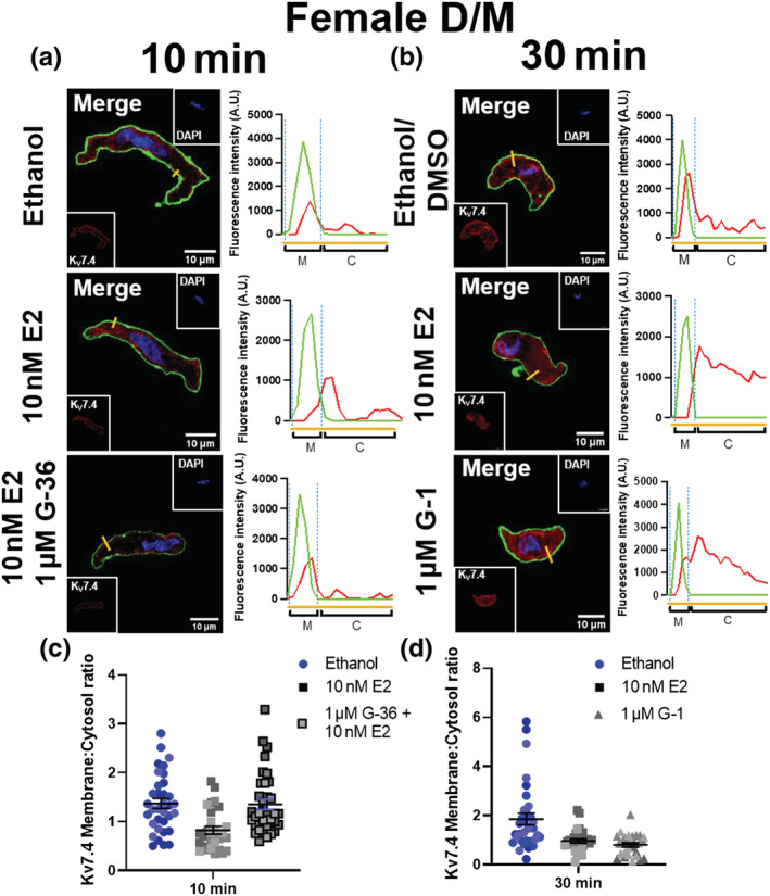
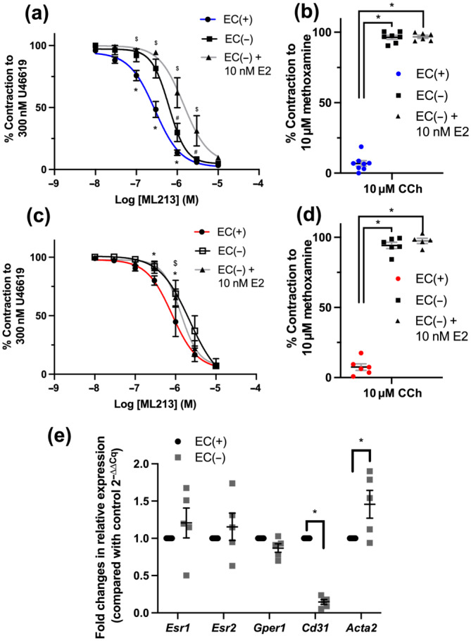
The K 7.2-5 activators S-1 and ML213 and the pan-K 7 inhibitor linopirdine were more effective in arteries from F-D/M compared with F-P/E animals. In VSMCs isolated from F-P/E rats, exploratory evidence indicates reduced membrane abundance of K 7.4 but not K 7.1, K 7.5 and Kcne4 when compared with cells from F-D/M. Plasma oestradiol was higher in F-P/E compared with F-D/M, and progesterone showed the converse pattern. Oestradiol/GPER1 agonist G-1 diminished K 7.4 encoded currents and ML213 relaxations and reduced the membrane abundance of K 7.4 and interaction between K 7.4 and heat shock protein 90 (HSP90), in arteries from F-D/M but not F-P/E.
GPER1 signalling decreased K 7.4 membrane abundance in conjunction with diminished interaction with HSP90, giving rise to a 'pro-contractile state'.
Kcnq 编码的 K7 通道(称为 K7.1-5)调节血管平滑肌细胞(VSMC)在静息时的收缩性以及作为受体介导反应的靶点。然而,目前的数据主要来自男性。考虑到性别、发情周期和性激素对血管反应性的已知影响,本研究对来自雌性 Wistar 大鼠发情周期(发情前期和发情期)和发情间期(发情前期和发情期)的肾和肠系膜动脉中的 K7 通道的分子和功能特性进行了特征描述。
在肾和肠系膜动脉中进行 RT-qPCR、免疫细胞化学、接近连接测定和线描肌描记法。通过液相色谱-串联质谱法测定循环性激素浓度。在表达与 G 蛋白偶联雌激素受体 1(GPER1)相关的 K7.4 通道的细胞上进行全细胞电生理学。
与发情前期和发情期的动物相比,K7.2-5 激活剂 S-1 和 ML213 以及泛 K7 抑制剂 linopirdine 在发情间期的动脉中更有效。与发情前期的大鼠相比,在发情前期的大鼠分离的 VSMCs 中,探索性证据表明 K7.4 的膜丰度降低,但 K7.1、K7.5 和 Kcne4 的膜丰度没有降低。与发情前期相比,发情前期的大鼠血浆雌二醇水平较高,孕酮水平则相反。雌二醇/GPER1 激动剂 G-1 减弱了 K7.4 编码的电流和 ML213 的松弛作用,并降低了发情前期动脉中 K7.4 的膜丰度和 K7.4 与热休克蛋白 90(HSP90)之间的相互作用,但对发情前期的动脉没有影响。
GPER1 信号降低了发情间期 K7.4 的膜丰度,同时降低了与 HSP90 的相互作用,导致出现“促收缩状态”。