Kennedy Gregory T, Azari Feredun S, Bernstein Elizabeth, Marfatia Isvita, Din Azra, Deshpande Charuhas, Galvis Nikki, Sorger Jonathan, Kucharczuk John C, Singhal Sunil
Department of Surgery, University of Pennsylvania Perelman School of Medicine, Philadelphia, PA, USA.
Department of Pathology, University of Pennsylvania Perelman School of Medicine, Philadelphia, PA, USA.
Transl Lung Cancer Res. 2022 Aug;11(8):1567-1577. doi: 10.21037/tlcr-21-1004.
Identifying ground glass opacities (GGOs) is challenging during robot-assisted thoracic surgery (RATS). Intraoperative molecular imaging (IMI) using tumor-targeted fluorescent tracers may address this clinical problem, but has never been evaluated in RATS. In a pilot study, we sought to determine whether IMI during RATS (RIMI) can localize GGOs.
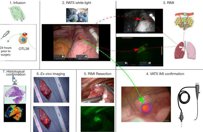
Ten patients with a cT1 GGO were enrolled. Prior to resection, participants received a folate-receptor targeted fluorescent tracer (OTL38). During RATS, a white-light robotic scope was utilized to identify tumors. RIMI was then conducted using a RATS thoracoscope with a wavelength-specific camera. Finally, a video-assisted thoracic surgery (VATS) thoracoscope designed to detect OTL38 was used as a control to compare to RIMI. The lesions were then resected under RIMI guidance.
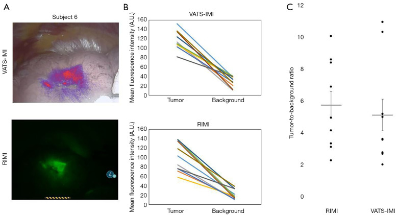
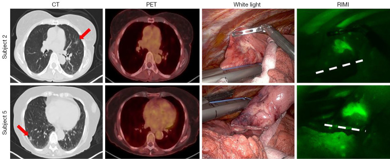
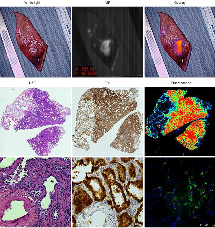
By white-light robotic scope, 7/10 (70%) GGOs were visually identifiable by pleuroparenchymal distortions. RIMI identified tumor-specific fluorescence in all (100%) subjects. RIMI clearly located the three nodules that could not be seen by robotic white-light imaging. The mean fluorescence intensity (MFI) of tumors was 99.48 arbitrary units (A.U.) (IQR, 75.72-130.49 A.U.), which was significantly higher than background tissue with mean MFI 20.61 A.U. (IQR, 13.49-29.93 A.U., P<0.0001). Mean signal-to-background ratio was 5.71 (range, 2.28-10.13). When compared to VATS-IMI as a control, there were no significant differences in MFI of tumors, background tissue, or signal-to-background ratios. In summary, RIMI compared favorably to VATS-IMI by all measured imaging characteristics.
RIMI is feasible for identification of GGOs during robotic resection as compared to white light thoracoscopy and compares favorably to VATS-IMI.
在机器人辅助胸外科手术(RATS)中识别磨玻璃影(GGO)具有挑战性。使用肿瘤靶向荧光示踪剂的术中分子成像(IMI)可能解决这一临床问题,但从未在RATS中进行过评估。在一项初步研究中,我们试图确定RATS期间的IMI(RIMI)是否能够定位GGO。
招募了10例cT1期GGO患者。在切除术前,参与者接受了一种叶酸受体靶向荧光示踪剂(OTL38)。在RATS手术期间,使用白光机器人胸腔镜识别肿瘤。然后使用带有波长特异性摄像头的RATS胸腔镜进行RIMI。最后,使用设计用于检测OTL38的电视辅助胸腔镜手术(VATS)胸腔镜作为对照与RIMI进行比较。然后在RIMI引导下切除病变。
通过白光机器人胸腔镜,7/10(70%)的GGO可通过胸膜实质扭曲在视觉上识别。RIMI在所有(100%)受试者中均识别出肿瘤特异性荧光。RIMI清楚地定位了机器人白光成像无法看到的三个结节。肿瘤的平均荧光强度(MFI)为99.48任意单位(A.U.)(四分位间距,75.72 - 130.49 A.U.),显著高于背景组织,背景组织的平均MFI为20.61 A.U.(四分位间距,13.49 - 29.93 A.U.,P<0.0001)。平均信号与背景比值为5.71(范围,2.28 - 10.13)。与作为对照的VATS-IMI相比,肿瘤、背景组织的MFI或信号与背景比值均无显著差异。总之,在所有测量的成像特征方面,RIMI与VATS-IMI相比表现良好。
与白光胸腔镜检查相比,RIMI在机器人切除术中识别GGO是可行的,并且与VATS-IMI相比表现良好。