Tamura Masanori, Furumatsu Takayuki, Hiranaka Takaaki, Kintaka Keisuke, Higashihara Naohiro, Kamatsuki Yusuke, Nakata Eiji, Ozaki Toshifumi
Department of Orthopaedic Surgery, Okayama University Graduate School of Medicine, Dentistry and Pharmaceutical Sciences, 2-5-1 Shikatacho, Kitaku, Okayama 700-8558, Japan.
Case Rep Orthop. 2022 Aug 31;2022:9776388. doi: 10.1155/2022/9776388. eCollection 2022.
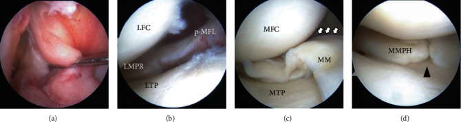
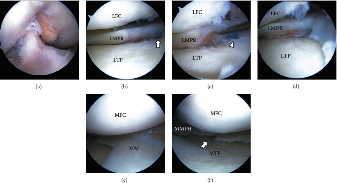
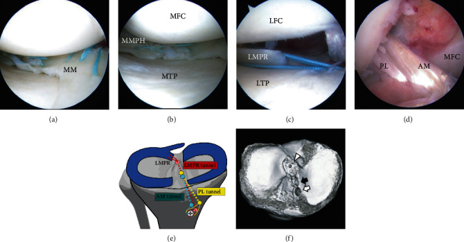
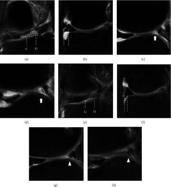
Lateral meniscus (LM) posterior root tear (LMPRT) is mainly caused by trauma, especially trauma associated with anterior cruciate ligament (ACL) injuries. Although a transtibial pullout repair or a side-to-side repair is commonly performed for LMPRT, to the best of our knowledge, there is no clinical report of LMPRT with tissue loss using the pullout technique. Thus, the purpose of this report was to describe a clinical, radiographic, and arthroscopic outcome after pullout repair for a case of LMPRT with a large defect with a chronic ACL tear and complex medial meniscus (MM) tears. A 31-year-old man complained of knee pain and restricted range of motion after twisting his knee when he stepped on an iron pipe. The patient had a football-related injury to his right knee 14 years before presentation, and since then, the patient's knee has given out more than 10 times but was left unassessed. Magnetic resonance imaging showed LMPRT with tissue loss, ACL tears, and complex MM tears. Transtibial pullout repair of the LMPRT with ACL reconstruction and MM repairs were performed. Following the pullout repair of the LMPRT, an approximately 6 mm gap remained between the LM posterior root and root insertion. However, magnetic resonance imaging and second-look arthroscopy at 1 year postoperatively revealed meniscal healing, gap filling with some regeneration tissue, of the LM posterior root. Furthermore, the lateral meniscus extrusion in the coronal plane improved from 3.1 mm (preoperative) to 1.6 mm (1 year postoperatively). Transtibial pullout repair with the remaining gap could be a viable treatment option for LMPRT with tissue loss, combined with ACL reconstruction.
外侧半月板(LM)后根部撕裂(LMPRT)主要由创伤引起,尤其是与前交叉韧带(ACL)损伤相关的创伤。尽管对于LMPRT通常进行经胫骨拉出修复或端端修复,但据我们所知,尚无使用拉出技术治疗伴有组织缺损的LMPRT的临床报告。因此,本报告的目的是描述一例伴有慢性ACL撕裂和复杂内侧半月板(MM)撕裂且存在大缺损的LMPRT患者经拉出修复后的临床、影像学和关节镜检查结果。一名31岁男性在踩到铁管时膝盖扭转后出现膝关节疼痛和活动范围受限。该患者在就诊前14年有与足球相关的右膝损伤,从那时起,患者的膝盖已出现超过10次打软腿情况,但未接受评估。磁共振成像显示存在LMPRT伴组织缺损、ACL撕裂和复杂的MM撕裂。对LMPRT进行了经胫骨拉出修复,并同时进行了ACL重建和MM修复。在LMPRT的拉出修复后,LM后根部与根部附着点之间仍存在约6毫米的间隙。然而,术后1年的磁共振成像和二次关节镜检查显示,LM后根部半月板愈合,间隙被一些再生组织填充。此外,冠状面的外侧半月板挤压从术前的3.1毫米改善至术后1年的1.6毫米。对于伴有组织缺损的LMPRT,经胫骨拉出修复并保留间隙,结合ACL重建,可能是一种可行的治疗选择。