Murata Takaaki, Ishimori Takahiro, Naitou Wataru, Igrashi Yuto, Suno Yuma, Kawachi Jun
Department of General Surgery, Shonan Kamakura General Hospital, Kanagawa, Japan.
Department of General Surgery, Shonan Kamakura General Hospital, Kanagawa, Japan.
Int J Surg Case Rep. 2022 Oct;99:107567. doi: 10.1016/j.ijscr.2022.107567. Epub 2022 Aug 31.
Ruptured extragastrointestinal stromal tumor (EGIST) are rare; therefore, there are no standard guidelines for its treatment. Herein, we report the successful laparoscopic resection of a ruptured EGIST.
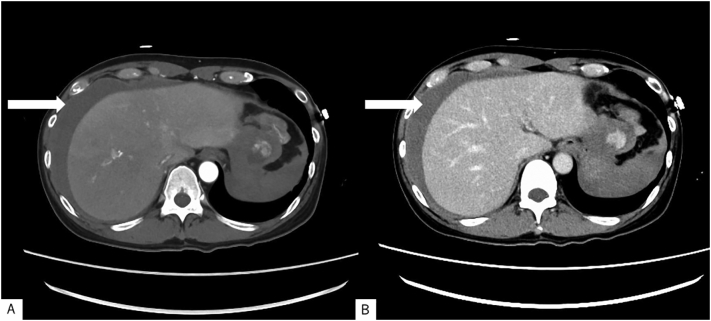
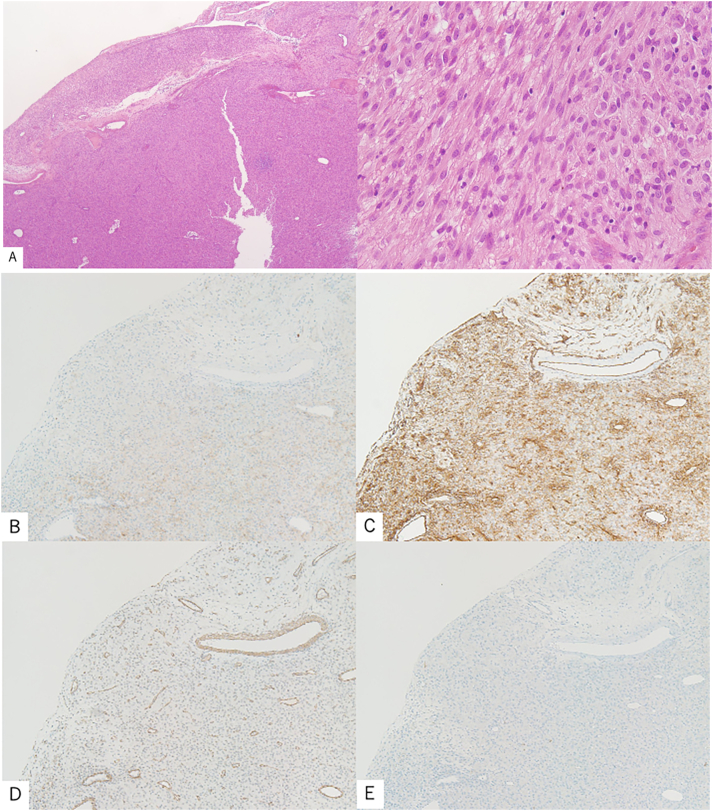
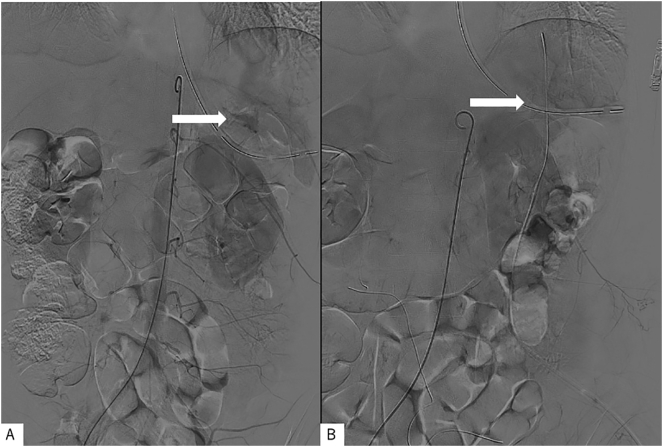
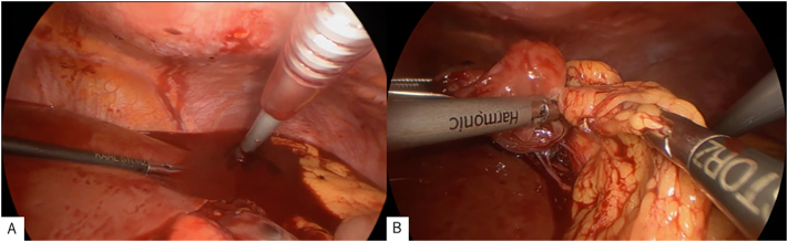
The patient was a 59-year-old man, a Jehovah's Witness, who presented with sudden onset of left-sided abdominal pain. Contrast-enhanced computed tomography (CECT) performed from a previous hospital revealed intra-abdominal hemorrhage. Repeat CECT at our institution revealed extravasation and serum ascites. A hematoma was found anterior to the omentum, and a tumor was detected which did not have continuity with the surrounding organs of the gastrointestinal tract. Complete tumor resection via laparoscopic surgery was performed and the specimen was sent for histopathology, which revealed bundle-like proliferation of spindle-shaped cells. Immunohistochemical staining was completed, which was positive for KIT and CD34. Based on surgical and pathological findings, the final diagnosis was extragastrointestinal stromal tumor originating from the omentum.
EGISTs have a similar morphology to that of gastrointestinal stromal tumors, but instead, arise outside the gastrointestinal tract. A significant differentiation and key to the diagnosis of EGIST is the absence of continuity with the gastrointestinal tract. The preferred treatment for EGIST is complete surgical resection, and the use of laparoscopy has not been well studied. Postoperative histopathological examination, along with immunohistochemical staining, aid confirmatory diagnosis.
Laparoscopic removal of EGISTs is a minimally invasive and potentially useful technique for the management of this tumor type.
胃肠道外间质瘤(EGIST)破裂罕见,因此尚无标准的治疗指南。在此,我们报告一例成功通过腹腔镜切除破裂EGIST的病例。
患者为一名59岁男性,耶和华见证会成员,突发左侧腹痛。外院的增强计算机断层扫描(CECT)显示腹腔内出血。我院重复CECT显示造影剂外渗和血清性腹水。在大网膜前方发现一个血肿,并检测到一个与胃肠道周围器官无连续性的肿瘤。通过腹腔镜手术完成了肿瘤完整切除,标本送病理检查,结果显示梭形细胞呈束状增生。完成免疫组化染色,结果显示KIT和CD34呈阳性。根据手术和病理结果,最终诊断为起源于大网膜的胃肠道外间质瘤。
EGIST的形态与胃肠道间质瘤相似,但起源于胃肠道外。EGIST的一个重要鉴别点和诊断关键是与胃肠道无连续性。EGIST的首选治疗方法是完整手术切除,腹腔镜的应用尚未得到充分研究。术后组织病理学检查及免疫组化染色有助于确诊。
腹腔镜切除EGIST是一种微创且可能有效的治疗该肿瘤类型的技术。