Department of Radiology, The Third Affiliated Hospital of Kunming Medical University, Kunming, 650018, China.
Department of Psychiatry, The Second Affiliated Hospital of Kunming Medical University, 374# DianMian Road, 650101, Kunming, China.
BMC Med Imaging. 2022 Sep 12;22(1):164. doi: 10.1186/s12880-022-00892-5.
Radiomics is an emerging image analysis framework that provides more details than conventional methods. In present study, we aimed to identify structural radiomics features of gray matter (GM) and white matter (WM), and to develop and validate the classification model for major depressive disorder (MDD) and subthreshold depression (StD) diagnosis using radiomics analysis.
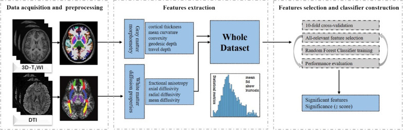
A consecutive cohort of 142 adolescents and young adults, including 43 cases with MDD, 49 cases with StD and 50 healthy controls (HC), were recruited and underwent the three-dimensional T1 weighted imaging (3D-TWI) and diffusion tensor imaging (DTI). We extracted radiomics features representing the shape and diffusion properties of GM and WM from all participants. Then, an all-relevant feature selection process embedded in a 10-fold cross-validation framework was used to identify features with significant power for discrimination. Random forest classifiers (RFC) were established and evaluated successively using identified features.
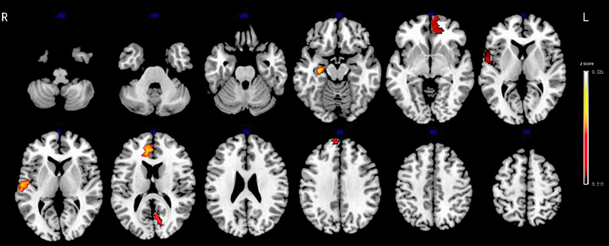
The results showed that a total of 3030 features were extracted after preprocessing, including 2262 shape-related features from each T1-weighted image representing GM morphometry and 768 features from each DTI representing the diffusion properties of WM. 25 features were selected ultimately, including ten features for MDD versus HC, eight features for StD versus HC, and seven features for MDD versus StD. The accuracies and area under curve (AUC) the RFC achieved were 86.75%, 0.93 for distinguishing MDD from HC with significant radiomics features located in the left medial orbitofrontal cortex, right superior and middle temporal regions, right anterior cingulate, left cuneus and hippocampus, 70.51%, 0.69 for discriminating StD from HC within left cuneus, medial orbitofrontal cortex, cerebellar vermis, hippocampus, anterior cingulate and amygdala, right superior and middle temporal regions, and 59.15%, 0.66 for differentiating MDD from StD within left medial orbitofrontal cortex, middle temporal and cuneus, right superior frontal, superior temporal regions and hippocampus, anterior cingulate, respectively.
These findings provide preliminary evidence that radiomics features of brain structure are valid for discriminating MDD and StD subjects from healthy controls. The MRI-based radiomics approach, with further improvement and validation, might be a potential facilitating method to clinical diagnosis of MDD or StD.
放射组学是一种新兴的图像分析框架,它提供的细节比传统方法更多。本研究旨在识别灰质(GM)和白质(WM)的结构放射组学特征,并使用放射组学分析开发和验证用于重度抑郁症(MDD)和阈下抑郁(StD)诊断的分类模型。
连续招募了 142 名青少年和年轻人,包括 43 例 MDD、49 例 StD 和 50 例健康对照组(HC),并对其进行了三维 T1 加权成像(3D-TWI)和扩散张量成像(DTI)。我们从所有参与者中提取了代表 GM 和 WM 形状和扩散特性的放射组学特征。然后,使用包含在 10 折交叉验证框架中的全相关特征选择过程来识别具有显著判别能力的特征。随后,使用鉴定的特征建立并评估随机森林分类器(RFC)。
预处理后共提取了 3030 个特征,包括来自每个 T1 加权图像的 2262 个代表 GM 形态学的形状相关特征和来自每个 DTI 的 768 个代表 WM 扩散特性的特征。最终选择了 25 个特征,包括 10 个用于 MDD 与 HC 的特征、8 个用于 StD 与 HC 的特征和 7 个用于 MDD 与 StD 的特征。RFC 达到的准确性和曲线下面积(AUC)分别为 86.75%,0.93 用于区分 MDD 与 HC,具有显著放射组学特征的区域位于左内侧眶额皮质、右额上和中颞叶、右前扣带回、左楔前叶和海马,70.51%,0.69 用于区分 StD 与 HC,位于左楔前叶、内侧眶额皮质、小脑蚓部、海马、前扣带回和杏仁核、右额上和中颞叶,59.15%,0.66 用于区分 MDD 与 StD,位于左内侧眶额皮质、中颞叶和楔前叶、右额上、中颞叶和海马、前扣带回、分别位于额上回和颞上回。
这些发现提供了初步证据,表明脑结构的放射组学特征可有效区分 MDD 和 StD 受试者与健康对照者。基于 MRI 的放射组学方法,通过进一步改进和验证,可能成为 MDD 或 StD 临床诊断的一种潜在辅助方法。