• 文献检索
  • 文档翻译
  • 深度研究
  • 学术资讯
  • Suppr Zotero 插件Zotero 插件
  • 邀请有礼
  • 套餐&价格
  • 历史记录
应用&插件
Suppr Zotero 插件Zotero 插件浏览器插件Mac 客户端Windows 客户端微信小程序
定价
高级版会员购买积分包购买API积分包
服务
文献检索文档翻译深度研究API 文档MCP 服务
关于我们
关于 Suppr公司介绍联系我们用户协议隐私条款
关注我们

Suppr 超能文献

核心技术专利:CN118964589B侵权必究
粤ICP备2023148730 号-1Suppr @ 2026

文献检索

告别复杂PubMed语法,用中文像聊天一样搜索,搜遍4000万医学文献。AI智能推荐,让科研检索更轻松。

立即免费搜索

文件翻译

保留排版,准确专业,支持PDF/Word/PPT等文件格式,支持 12+语言互译。

免费翻译文档

深度研究

AI帮你快速写综述,25分钟生成高质量综述,智能提取关键信息,辅助科研写作。

立即免费体验

多发性硬化症的脑血管反应性在免疫调节过程中炎症减轻后得到恢复。

Cerebrovascular reactivity in multiple sclerosis is restored with reduced inflammation during immunomodulation.

机构信息

Department of Neuroscience, Imaging, and Clinical Sciences, University G. d'Annunzio of Chieti-Pescara, Chieti, Italy.

Institute for Advanced Biomedical Technologies (ITAB), University G. d'Annunzio of Chieti-Pescara, Chieti, Italy.

出版信息

Sci Rep. 2022 Sep 14;12(1):15453. doi: 10.1038/s41598-022-19113-8.

DOI:10.1038/s41598-022-19113-8
PMID:36104366
原文链接:https://pmc.ncbi.nlm.nih.gov/articles/PMC9474533/
Abstract

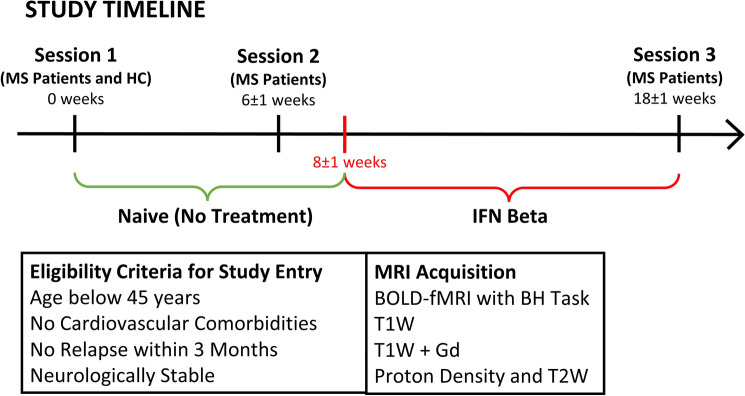
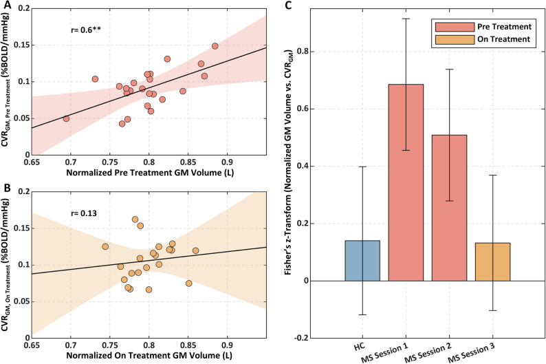
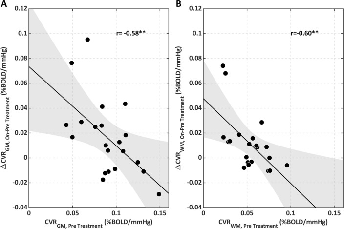
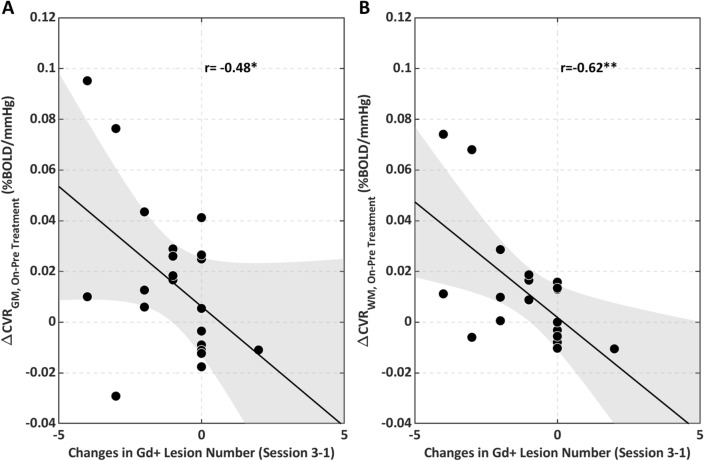
Cerebrovascular reactivity (CVR) reflects the capacity of the brain's vasculature to increase blood flow following a vasodilatory stimulus. Reactivity is an essential property of the brain's blood vessels that maintains nutrient supplies in the face of changing demand. In Multiple Sclerosis (MS), CVR may be diminished with brain inflammation and this may contribute to neurodegeneration. We test the hypothesis that CVR is altered with MS neuroinflammation and that it is restored when inflammation is reduced. Using a breath-hold task during functional Magnetic Resonance Imaging (MRI), we mapped grey matter and white matter CVRs (CVR and CVR, respectively) in 23 young MS patients, eligible for disease modifying therapy, before and during Interferon beta treatment. Inflammatory activity was inferred from the presence of Gadolinium enhancing lesions at MRI. Eighteen age and gender-matched healthy controls (HC) were also assessed. Enhancing lesions were observed in 12 patients at the start of the study and in 3 patients during treatment. Patients had lower pre-treatment CVR (p = 0.04) and CVR (p = 0.02) compared to HC. In patients, a lower pre-treatment CVR was associated with a lower GM volume (r = 0.60, p = 0.003). On-treatment, there was an increase in CVR (p = 0.02) and CVR (p = 0.03) that negatively correlated with pre-treatment CVR (GM: r = - 0.58, p = 0.005; WM: r = - 0.60, p = 0.003). CVR increased when enhancing lesions reduced in number (GM: r = - 0.48, p = 0.02, WM: r = - 0.62, p = 0.003). Resolution of inflammation may restore altered cerebrovascular function limiting neurodegeneration in MS. Imaging of cerebrovascular function may thereby inform tissue physiology and improve treatment monitoring.

摘要

脑血管反应性(CVR)反映了大脑血管在接受血管扩张刺激后增加血流量的能力。反应性是大脑血管的基本特性,可在需求变化时维持营养供应。在多发性硬化症(MS)中,脑炎症可能会降低 CVR,这可能导致神经退行性变。我们假设 CVR 在 MS 神经炎症中发生改变,并且在炎症减轻时恢复。在功能磁共振成像(MRI)期间使用屏气任务,我们在 23 名年轻的多发性硬化症患者中分别映射了灰质和白质 CVR(CVR 和 CVR),这些患者符合疾病修饰治疗的条件,分别在干扰素β治疗之前和期间进行。通过 MRI 中的钆增强病变来推断炎症活动。还评估了 18 名年龄和性别匹配的健康对照者(HC)。在研究开始时,12 名患者和治疗期间的 3 名患者观察到增强病变。与 HC 相比,患者的治疗前 CVR(p = 0.04)和 CVR(p = 0.02)较低。在患者中,较低的治疗前 CVR 与较低的 GM 体积相关(r = 0.60,p = 0.003)。在治疗期间,CVR(p = 0.02)和 CVR(p = 0.03)增加,与治疗前 CVR 呈负相关(GM:r = -0.58,p = 0.005;WM:r = -0.60,p = 0.003)。当增强病变数量减少时,CVR 增加(GM:r = -0.48,p = 0.02,WM:r = -0.62,p = 0.003)。炎症的解决可能会恢复改变的脑血管功能,从而限制 MS 中的神经退行性变。脑血管功能成像因此可以提供组织生理学信息并改善治疗监测。

https://cdn.ncbi.nlm.nih.gov/pmc/blobs/f917/9474533/5dfb7bc5e6eb/41598_2022_19113_Fig7_HTML.jpg
https://cdn.ncbi.nlm.nih.gov/pmc/blobs/f917/9474533/c6f1c2b1f0e2/41598_2022_19113_Fig1_HTML.jpg
https://cdn.ncbi.nlm.nih.gov/pmc/blobs/f917/9474533/3efd094f7763/41598_2022_19113_Fig2_HTML.jpg
https://cdn.ncbi.nlm.nih.gov/pmc/blobs/f917/9474533/298386967b5e/41598_2022_19113_Fig3_HTML.jpg
https://cdn.ncbi.nlm.nih.gov/pmc/blobs/f917/9474533/3e4e2e9bf412/41598_2022_19113_Fig4_HTML.jpg
https://cdn.ncbi.nlm.nih.gov/pmc/blobs/f917/9474533/94aa06702394/41598_2022_19113_Fig5_HTML.jpg
https://cdn.ncbi.nlm.nih.gov/pmc/blobs/f917/9474533/1f972d354d20/41598_2022_19113_Fig6_HTML.jpg
https://cdn.ncbi.nlm.nih.gov/pmc/blobs/f917/9474533/5dfb7bc5e6eb/41598_2022_19113_Fig7_HTML.jpg

相似文献

1
Cerebrovascular reactivity in multiple sclerosis is restored with reduced inflammation during immunomodulation.多发性硬化症的脑血管反应性在免疫调节过程中炎症减轻后得到恢复。
Sci Rep. 2022 Sep 14;12(1):15453. doi: 10.1038/s41598-022-19113-8.
2
Cerebrovascular reactivity and its correlation with age in patients with multiple sclerosis.多发性硬化症患者的脑血管反应性及其与年龄的相关性。
Brain Imaging Behav. 2020 Oct;14(5):1889-1898. doi: 10.1007/s11682-019-00132-5.
3
Impaired cerebrovascular reactivity in multiple sclerosis.多发性硬化症中脑血管反应性受损。
JAMA Neurol. 2014 Oct;71(10):1275-81. doi: 10.1001/jamaneurol.2014.1668.
4
Cerebrovascular reactivity and disease activity in relapsing-remitting multiple sclerosis.复发缓解型多发性硬化症中的脑血管反应性与疾病活动度。
Adv Clin Exp Med. 2020 Feb;29(2):183-188. doi: 10.17219/acem/114762.
5
Temporal Profile of Cerebrovascular Reactivity Impairment, Gray Matter Volumes, and Persistent Symptoms after Mild Traumatic Head Injury.轻度创伤性脑损伤后脑血管反应性损害、灰质体积和持续症状的时间特征
Front Neurol. 2016 May 11;7:70. doi: 10.3389/fneur.2016.00070. eCollection 2016.
6
Developmental trajectories of cerebrovascular reactivity in healthy children and young adults assessed with magnetic resonance imaging.使用磁共振成像评估健康儿童和年轻人脑血管反应性的发育轨迹。
J Physiol. 2016 May 15;594(10):2681-9. doi: 10.1113/JP271056. Epub 2016 Mar 17.
7
Quantifying cerebrovascular reactivity in anterior and posterior cerebral circulations during voluntary breath holding.在自主屏气期间量化大脑前、后循环中的脑血管反应性。
Exp Physiol. 2016 Dec 1;101(12):1517-1527. doi: 10.1113/EP085764.
8
A Promising Subject-Level Classification Model for Acute Concussion Based on Cerebrovascular Reactivity Metrics.一种基于脑血管反应性指标的急性脑震荡有前景的受试者水平分类模型。
J Neurotrauma. 2021 Apr 15;38(8):1036-1047. doi: 10.1089/neu.2020.7272. Epub 2020 Dec 14.
9
Functional Neurosonology Reveals Impaired Cerebrovascular Reactivity in Multiple Sclerosis.功能神经超声揭示多发性硬化症患者脑血管反应受损。
J Neuroimaging. 2019 Sep;29(5):589-591. doi: 10.1111/jon.12617. Epub 2019 Apr 16.
10
Characterization of gray-matter multiple sclerosis lesions using double inversion recovery, diffusion, contrast-enhanced, and volumetric MRI.采用双反转恢复、弥散、对比增强和容积 MRI 对灰质多发性硬化病变进行特征描述。
Mult Scler Relat Disord. 2019 Jun;31:74-81. doi: 10.1016/j.msard.2019.03.021. Epub 2019 Mar 29.

引用本文的文献

1
Quantitative mapping of cerebrovascular reactivity amplitude and delay with breath-hold BOLD fMRI when end-tidal CO quality is low.当呼气末二氧化碳质量较低时,利用屏气功能磁共振成像对脑血管反应性幅度和延迟进行定量映射。
Imaging Neurosci (Camb). 2025 Apr 8;3. doi: 10.1162/imag_a_00536. eCollection 2025.
2
Differentiation of MS lesions through analysis of microvascular distribution.通过微血管分布分析对多发性硬化症病变进行鉴别
Imaging Neurosci (Camb). 2024 Nov 8;2. doi: 10.1162/imag_a_00357. eCollection 2024.
3
Quantitative mapping of cerebrovascular reactivity amplitude and delay with breath-hold BOLD fMRI when end-tidal CO quality is low.

本文引用的文献

1
Cerebral blood flow dependency on systemic arterial circulation in progressive multiple sclerosis.进展性多发性硬化症中脑血流对全身动脉循环的依赖性。
Eur Radiol. 2022 Sep;32(9):6468-6479. doi: 10.1007/s00330-022-08731-5. Epub 2022 Apr 1.
2
Cerebrovascular reactivity and disease activity in relapsing-remitting multiple sclerosis.复发缓解型多发性硬化症中的脑血管反应性与疾病活动度。
Adv Clin Exp Med. 2020 Feb;29(2):183-188. doi: 10.17219/acem/114762.
3
A roadmap to precision medicine for multiple sclerosis.多发性硬化症精准医学路线图。
当呼气末二氧化碳质量较低时,利用屏气功能磁共振成像对脑血管反应性幅度和延迟进行定量映射。
bioRxiv. 2025 Mar 15:2024.11.18.624159. doi: 10.1101/2024.11.18.624159.
Mult Scler. 2020 Apr;26(5):522-532. doi: 10.1177/1352458519881558. Epub 2020 Jan 22.
4
The role of brain vasculature in neurodegenerative disorders.脑血管在神经退行性疾病中的作用。
Nat Neurosci. 2018 Oct;21(10):1318-1331. doi: 10.1038/s41593-018-0234-x. Epub 2018 Sep 24.
5
The role of the cerebellum in multiple sclerosis-150 years after Charcot.小脑在多发性硬化症中的作用——夏科 150 年后的研究。
Neurosci Biobehav Rev. 2018 Jun;89:85-98. doi: 10.1016/j.neubiorev.2018.02.012. Epub 2018 Feb 23.
6
Vascular pathology in multiple sclerosis: reframing pathogenesis around the blood-brain barrier.多发性硬化中的血管病理学:围绕血脑屏障重新构建发病机制。
J Neurol Neurosurg Psychiatry. 2018 Jan;89(1):42-52. doi: 10.1136/jnnp-2017-316011. Epub 2017 Aug 31.
7
Cerebral blood flow modulation insufficiency in brain networks in multiple sclerosis: A hypercapnia MRI study.多发性硬化症脑网络中脑血流调节功能不全:一项高碳酸血症MRI研究。
J Cereb Blood Flow Metab. 2016 Dec;36(12):2087-2095. doi: 10.1177/0271678X16654922. Epub 2016 Jun 15.
8
The topograpy of demyelination and neurodegeneration in the multiple sclerosis brain.多发性硬化症脑部脱髓鞘和神经变性的拓扑结构。
Brain. 2016 Mar;139(Pt 3):807-15. doi: 10.1093/brain/awv398. Epub 2016 Feb 8.
9
Cerebral hypoperfusion: a new pathophysiologic concept in multiple sclerosis?脑灌注不足:多发性硬化症中的一个新的病理生理概念?
J Cereb Blood Flow Metab. 2015 Sep;35(9):1406-10. doi: 10.1038/jcbfm.2015.131. Epub 2015 Jun 24.
10
Increased perfusion in normal appearing white matter in high inflammatory multiple sclerosis patients.高炎症多发性硬化症患者正常外观白质灌注增加。
PLoS One. 2015 Mar 16;10(3):e0119356. doi: 10.1371/journal.pone.0119356. eCollection 2015.