• 文献检索
  • 文档翻译
  • 深度研究
  • 学术资讯
  • Suppr Zotero 插件Zotero 插件
  • 邀请有礼
  • 套餐&价格
  • 历史记录
应用&插件
Suppr Zotero 插件Zotero 插件浏览器插件Mac 客户端Windows 客户端微信小程序
定价
高级版会员购买积分包购买API积分包
服务
文献检索文档翻译深度研究API 文档MCP 服务
关于我们
关于 Suppr公司介绍联系我们用户协议隐私条款
关注我们

Suppr 超能文献

核心技术专利:CN118964589B侵权必究
粤ICP备2023148730 号-1Suppr @ 2026

文献检索

告别复杂PubMed语法,用中文像聊天一样搜索,搜遍4000万医学文献。AI智能推荐,让科研检索更轻松。

立即免费搜索

文件翻译

保留排版,准确专业,支持PDF/Word/PPT等文件格式,支持 12+语言互译。

免费翻译文档

深度研究

AI帮你快速写综述,25分钟生成高质量综述,智能提取关键信息,辅助科研写作。

立即免费体验

视网膜内皮及眼部其他非肌肉组织中的收缩蛋白。

Contractile proteins in retinal endothelium and other non-muscle tissues of the eye.

作者信息

Rahi A, Ashton N

出版信息

Br J Ophthalmol. 1978 Sep;62(9):627-43. doi: 10.1136/bjo.62.9.627.

DOI:10.1136/bjo.62.9.627
PMID:361070
原文链接:https://pmc.ncbi.nlm.nih.gov/articles/PMC1043310/
Abstract

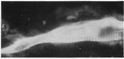
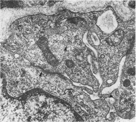
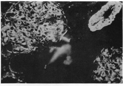
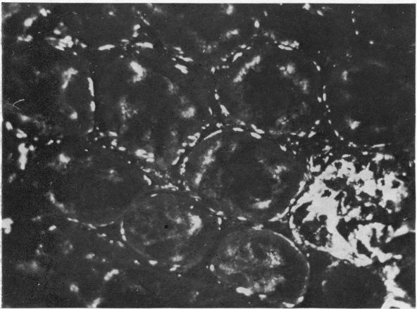
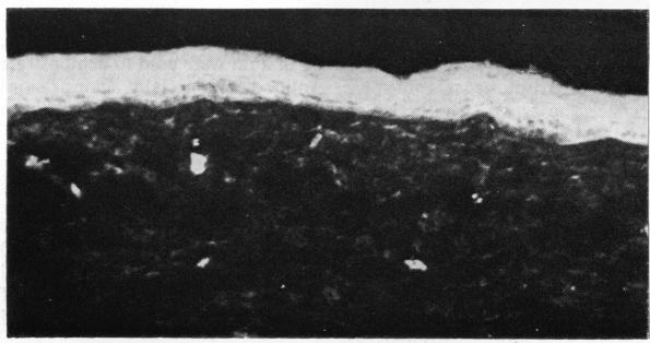
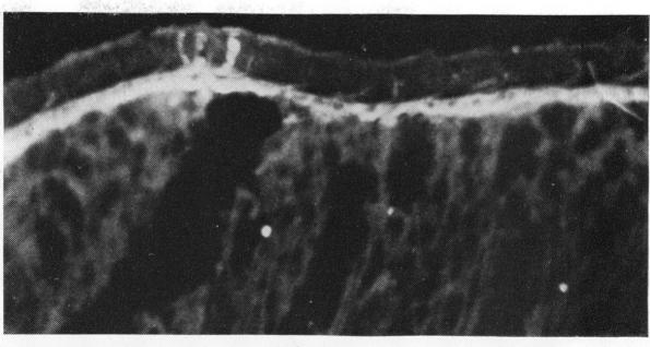
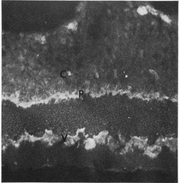
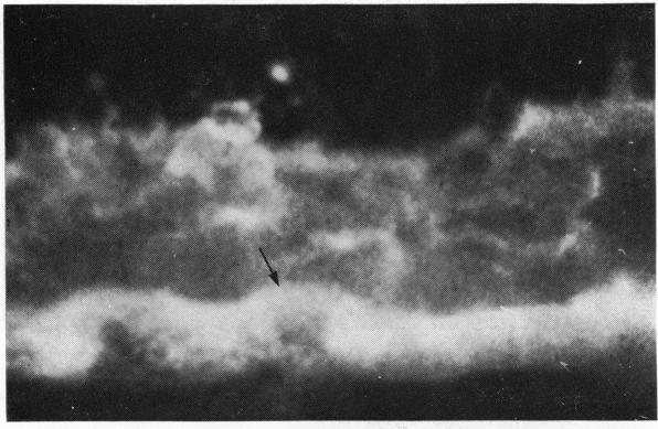
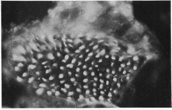
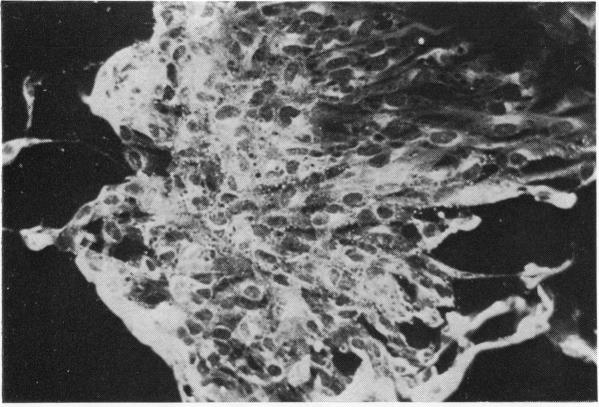
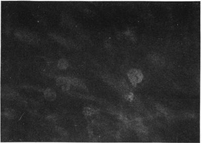
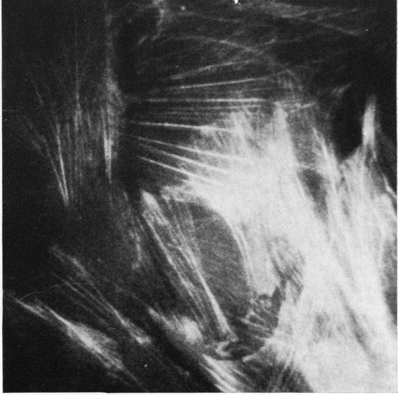
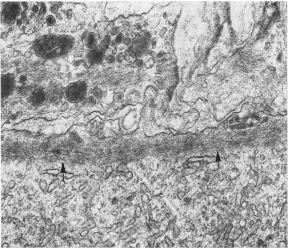
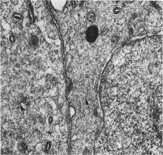
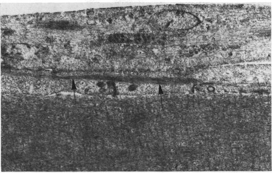
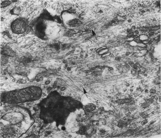
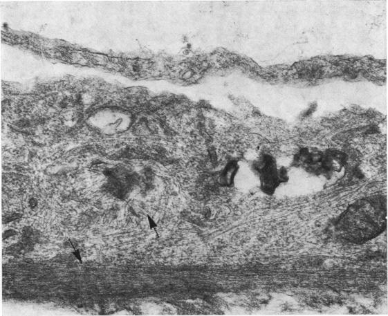
The present study is the first investigation to demonstrate, by employing the combined approach of immunologically and electron microscope methods, the presence of actin-like contractile proteins in the mammalian retina, the corneal epithelium and endothelium, the iris, and the ciliary body, and to confirm their presence in lens epithelium. This is also the first report to demonstrate by these methods the presence of microfilaments and intermediate filaments in retinal vascular endothelium. Since we have shown that actin filaments are especially abundant in immature retinal endothelial cells, the question of their function arises, and we have discussed their possible relevance to the closure of immature retinal vessels when exposed to hyperoxia.

摘要

本研究首次采用免疫和电子显微镜相结合的方法,证实了肌动蛋白样收缩蛋白在哺乳动物视网膜、角膜上皮和内皮、虹膜及睫状体中的存在,并进一步确认其在晶状体上皮中的存在。这也是首次通过这些方法证实视网膜血管内皮中存在微丝和中间丝。鉴于我们已表明肌动蛋白丝在未成熟视网膜内皮细胞中尤为丰富,其功能问题随之而来,我们还讨论了在高氧环境下,它们与未成熟视网膜血管闭合的可能关联。

https://cdn.ncbi.nlm.nih.gov/pmc/blobs/4cd0/1043310/f38069865c84/brjopthal00225-0065-a.jpg
https://cdn.ncbi.nlm.nih.gov/pmc/blobs/4cd0/1043310/e95f4f4aeb39/brjopthal00225-0051-a.jpg
https://cdn.ncbi.nlm.nih.gov/pmc/blobs/4cd0/1043310/9ea63c4f693e/brjopthal00225-0051-b.jpg
https://cdn.ncbi.nlm.nih.gov/pmc/blobs/4cd0/1043310/66f3e7ea5a30/brjopthal00225-0051-c.jpg
https://cdn.ncbi.nlm.nih.gov/pmc/blobs/4cd0/1043310/0f907d56e4fa/brjopthal00225-0052-a.jpg
https://cdn.ncbi.nlm.nih.gov/pmc/blobs/4cd0/1043310/9e4a9892518b/brjopthal00225-0052-b.jpg
https://cdn.ncbi.nlm.nih.gov/pmc/blobs/4cd0/1043310/7df22e0f5369/brjopthal00225-0053-a.jpg
https://cdn.ncbi.nlm.nih.gov/pmc/blobs/4cd0/1043310/524be7f703d5/brjopthal00225-0053-b.jpg
https://cdn.ncbi.nlm.nih.gov/pmc/blobs/4cd0/1043310/017cd9126508/brjopthal00225-0053-c.jpg
https://cdn.ncbi.nlm.nih.gov/pmc/blobs/4cd0/1043310/a215e786a646/brjopthal00225-0054-a.jpg
https://cdn.ncbi.nlm.nih.gov/pmc/blobs/4cd0/1043310/ef183716a625/brjopthal00225-0054-b.jpg
https://cdn.ncbi.nlm.nih.gov/pmc/blobs/4cd0/1043310/e9d5288bd7b5/brjopthal00225-0055-a.jpg
https://cdn.ncbi.nlm.nih.gov/pmc/blobs/4cd0/1043310/0a527d22ea1a/brjopthal00225-0055-b.jpg
https://cdn.ncbi.nlm.nih.gov/pmc/blobs/4cd0/1043310/d2a8045f74de/brjopthal00225-0055-c.jpg
https://cdn.ncbi.nlm.nih.gov/pmc/blobs/4cd0/1043310/91fc4103437b/brjopthal00225-0056-a.jpg
https://cdn.ncbi.nlm.nih.gov/pmc/blobs/4cd0/1043310/c6fa9fcd2d6f/brjopthal00225-0056-b.jpg
https://cdn.ncbi.nlm.nih.gov/pmc/blobs/4cd0/1043310/f9aef4c0fbc9/brjopthal00225-0057-a.jpg
https://cdn.ncbi.nlm.nih.gov/pmc/blobs/4cd0/1043310/77f84f032172/brjopthal00225-0057-b.jpg
https://cdn.ncbi.nlm.nih.gov/pmc/blobs/4cd0/1043310/365f14f752b0/brjopthal00225-0058-a.jpg
https://cdn.ncbi.nlm.nih.gov/pmc/blobs/4cd0/1043310/81f569d145cb/brjopthal00225-0058-b.jpg
https://cdn.ncbi.nlm.nih.gov/pmc/blobs/4cd0/1043310/e8d2bc01b2db/brjopthal00225-0059-a.jpg
https://cdn.ncbi.nlm.nih.gov/pmc/blobs/4cd0/1043310/a0cb15b02a93/brjopthal00225-0060-a.jpg
https://cdn.ncbi.nlm.nih.gov/pmc/blobs/4cd0/1043310/ad7d14764ec4/brjopthal00225-0061-a.jpg
https://cdn.ncbi.nlm.nih.gov/pmc/blobs/4cd0/1043310/030fe5875708/brjopthal00225-0062-a.jpg
https://cdn.ncbi.nlm.nih.gov/pmc/blobs/4cd0/1043310/1b73334ababa/brjopthal00225-0063-a.jpg
https://cdn.ncbi.nlm.nih.gov/pmc/blobs/4cd0/1043310/31153c8cd5b1/brjopthal00225-0064-a.jpg
https://cdn.ncbi.nlm.nih.gov/pmc/blobs/4cd0/1043310/f38069865c84/brjopthal00225-0065-a.jpg

相似文献

1
Contractile proteins in retinal endothelium and other non-muscle tissues of the eye.视网膜内皮及眼部其他非肌肉组织中的收缩蛋白。
Br J Ophthalmol. 1978 Sep;62(9):627-43. doi: 10.1136/bjo.62.9.627.
2
Actin filaments in retinal pericytes and endothelial cells.视网膜周细胞和内皮细胞中的肌动蛋白丝。
Invest Ophthalmol Vis Sci. 1980 Dec;19(12):1433-41.
3
Contractile proteins in ocular tissues. Their role in health and disease.
Trans Ophthalmol Soc U K (1962). 1980 Apr;100(Pt 1):155-61.
4
In situ demonstration of actin in normal and injured ocular tissues using 7-nitrobenz-2-oxa-1,3-diazole phallacidin.使用7-硝基苯并-2-恶唑-1,3-二氮杂茂鬼笔环肽对正常和损伤眼组织中的肌动蛋白进行原位示踪
Cell Motil. 1982;2(4):343-54. doi: 10.1002/cm.970020404.
5
[Histological changes in the corneal endothelium, retina and pigment epithelium of the eye of rabbits after vitrectomy using ultrasonic phacofragmentators].[使用超声乳化仪进行玻璃体切除术后兔眼角膜内皮、视网膜及色素上皮的组织学变化]
Vestn Oftalmol. 1985 Nov-Dec;101(6):27-31.
6
Characterization of intermediate filaments and their structural organization during epithelium formation in pigmented epithelial cells of the retina in vitro.体外培养的视网膜色素上皮细胞上皮形成过程中中间丝的特征及其结构组织
Cell Tissue Res. 1986;244(1):87-93. doi: 10.1007/BF00218385.
7
Organization and function of stress fibers in cells in vitro and in situ. A review.体外和原位细胞中应力纤维的组织与功能。综述
Cell Muscle Motil. 1984;5:83-137. doi: 10.1007/978-1-4684-4592-3_2.
8
Actin filament localization and distribution in the young adult mouse cornea: a correlative immunofluorescent and cytochemical study.成年小鼠角膜中肌动蛋白丝的定位与分布:一项免疫荧光与细胞化学相关研究
Exp Eye Res. 1983 Feb;36(2):171-80. doi: 10.1016/0014-4835(83)90003-9.
9
[Immunohistochemical localization of myosin and actin in various nonmuscle cell types of rat eyes].
Verh Anat Ges. 1978(72):279-82.
10
Epithelialization of the corneal endothelium in posterior polymorphous dystrophy.
Invest Ophthalmol Vis Sci. 1980 Jul;19(7):832-5.

引用本文的文献

1
Tumours of the anterior uvea. III. Oxytalan fibres in the differential diagnosis of leiomyoma and malignant melanoma of the iris.前葡萄膜肿瘤。III. 弹力纤维在虹膜平滑肌瘤与恶性黑色素瘤鉴别诊断中的作用
Br J Ophthalmol. 1980 Nov;64(11):867-74. doi: 10.1136/bjo.64.11.867.
2
Structural basis of contraction in vitreal fibrous membranes.玻璃体纤维膜收缩的结构基础。
Br J Ophthalmol. 1981 Nov;65(11):737-49. doi: 10.1136/bjo.65.11.737.
3
The localization of actin in dividing corneal endothelial cells demonstrated with nitrobenzoxadiazole phallacidin.

本文引用的文献

1
Neovascularization in ocular disease.眼部疾病中的新生血管形成。
Trans Ophthalmol Soc U K (1962). 1961;81:145-61.
2
Effect of oxygen on developing retinal vessels with particular reference to the problem of retrolental fibroplasia.氧气对发育中的视网膜血管的影响,尤其涉及晶状体后纤维增生问题。
Br J Ophthalmol. 1954 Jul;38(7):397-432. doi: 10.1136/bjo.38.7.397.
3
Endothelial contraction induced by histamine-type mediators: an electron microscopic study.组胺类介质诱导的内皮细胞收缩:一项电子显微镜研究。
用硝基苯并恶二唑鬼笔环肽显示肌动蛋白在分裂角膜内皮细胞中的定位。
Cell Tissue Res. 1983;229(3):533-9. doi: 10.1007/BF00207696.
4
Microfilaments in the cells of the human trabecular meshwork.
Br J Ophthalmol. 1979 Jan;63(1):3-8. doi: 10.1136/bjo.63.1.3.
J Cell Biol. 1969 Sep;42(3):647-72. doi: 10.1083/jcb.42.3.647.
4
Microtubules in the lens.晶状体中的微管。
Arch Ophthalmol. 1968 Feb;79(2):189-95. doi: 10.1001/archopht.1968.03850040191017.
5
Microfilaments in cellular and developmental processes.细胞与发育过程中的微丝。
Science. 1971 Jan 15;171(3967):135-43. doi: 10.1126/science.171.3967.135.
6
The ultrastructure of mammalian arterioles and precapillary sphincters.哺乳动物小动脉和毛细血管前括约肌的超微结构。
J Ultrastruct Res. 1967 Apr;18(1):181-223. doi: 10.1016/s0022-5320(67)80239-9.
7
Ultrastructural features of cytofilaments within mammalian endothelial cells.哺乳动物内皮细胞内细胞丝的超微结构特征
Z Zellforsch Mikrosk Anat. 1967;83(1):40-8. doi: 10.1007/BF00334737.
8
Cell elongation in the cultured embryonic chick lens epithelium with and without protein synthesis. Involvement of microtubules.有或无蛋白质合成情况下培养的鸡胚晶状体上皮细胞的伸长。微管的作用。
J Cell Biol. 1972 Oct;55(1):82-92. doi: 10.1083/jcb.55.1.82.
9
Effect of oxygen on the developing retinal vessels of the rabbit. IV. Effect of hyperoxia on rabbit retinal vessels in tissue culture.氧对家兔视网膜血管发育的影响。IV. 高氧对组织培养中家兔视网膜血管的影响。
Exp Eye Res. 1974 Nov;19(5):449-75. doi: 10.1016/0014-4835(74)90053-0.
10
An electron-microscope study of adult mouse lens: some ultrastructural specializations.
J Ultrastruct Res. 1974 Feb;46(2):239-53. doi: 10.1016/s0022-5320(74)80059-6.