Grierson I, Rahi A H
Br J Ophthalmol. 1981 Nov;65(11):737-49. doi: 10.1136/bjo.65.11.737.
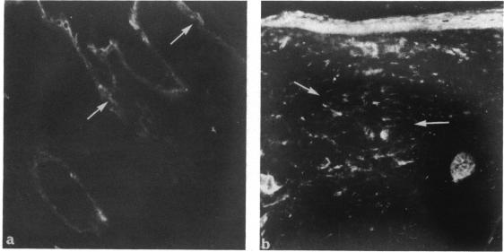
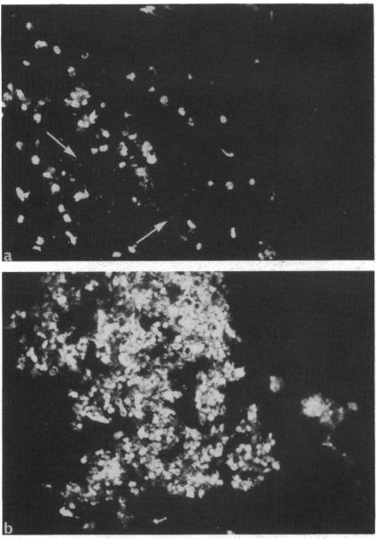
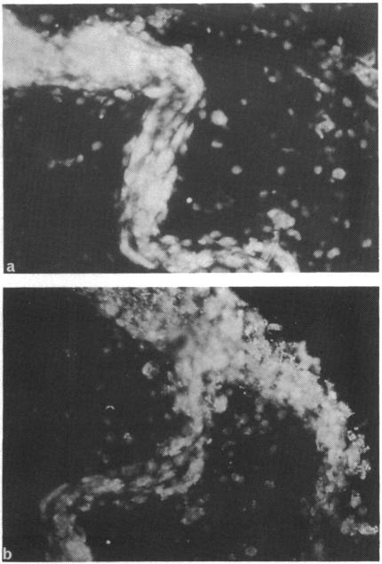
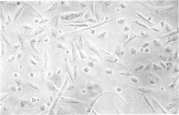
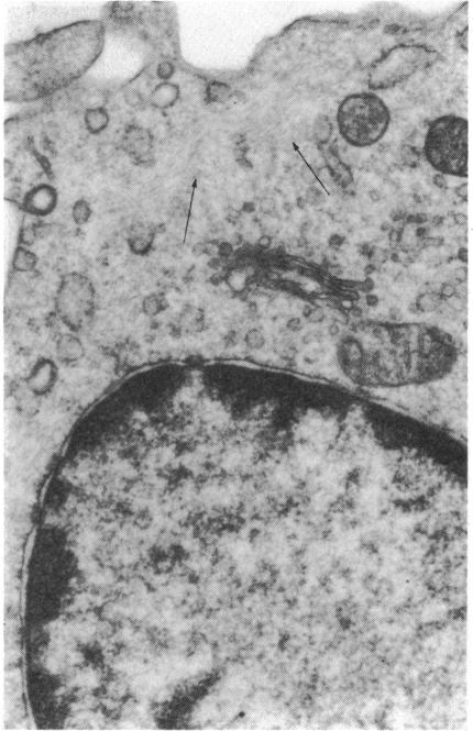
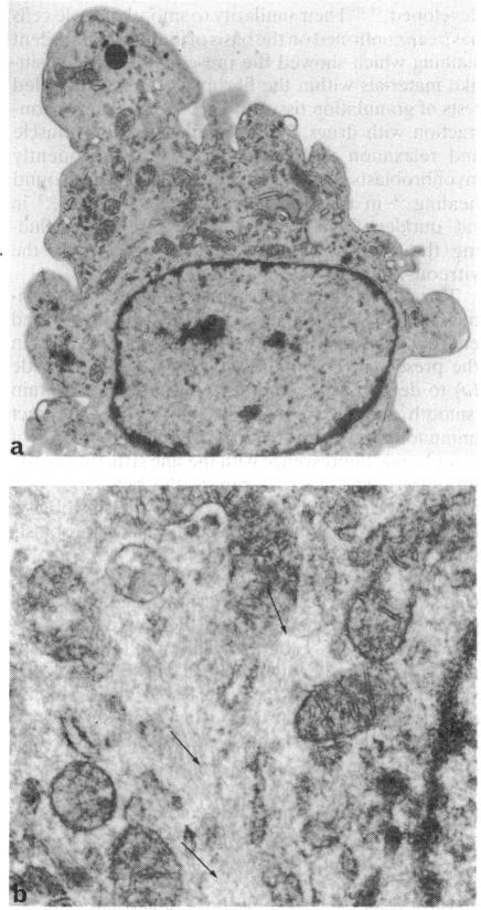
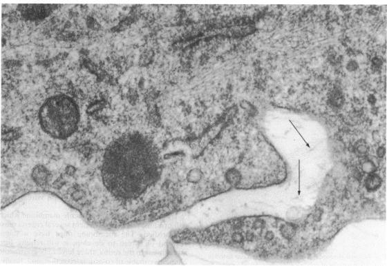
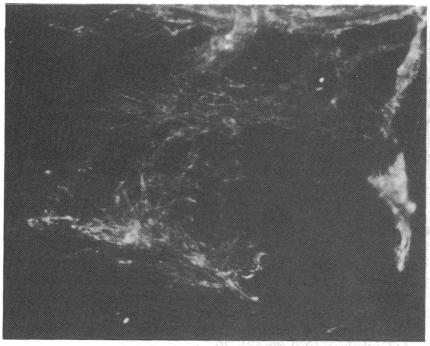
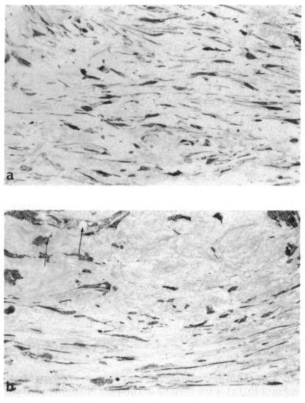
A combined ultrastructural and immunofluorescent study was conducted on experimentally induced fibrous membranes in the vitreous of adult rabbits. Autochthonous tissue cultured fibroblasts were injected into the mid-vitreous of one eye of each of 25 rabbits. The animals were monitored routinely with an ophthalmoscope and slit-lamp and were killed at various time periods between 5 minutes and 6 months. Appropriate tissue was taken for light microscopy, transmission electron microscopy, scanning electron microscopy, and indirect immunofluorescence. With this model we were able to show that the contractile elements in fibrous membranes are probably modified fibroblasts called myofibroblasts which are most abundant 3 to 6 weeks after injection. This is the time when retinal detachment usually occurs. It is our impression that, as traction membranes develop, there is not so much an increase in the contractile elements of the constituent cells as a rearrangement of the existing cytoplasmic microfilaments into compact highly organised bundles called stress cables. The behaviour and ultrastructural characteristics of intravitreal fibroblasts compare with the action of fibroblasts in the healing of wounds.
对成年兔玻璃体内实验性诱导形成的纤维膜进行了超微结构和免疫荧光联合研究。将自体组织培养的成纤维细胞注入25只兔子每只眼睛的玻璃体中部。定期用检眼镜和裂隙灯对动物进行监测,并在5分钟至6个月的不同时间段处死。取适当的组织进行光学显微镜、透射电子显微镜、扫描电子显微镜和间接免疫荧光检查。利用该模型,我们能够证明纤维膜中的收缩元件可能是被称为肌成纤维细胞的修饰成纤维细胞,在注射后3至6周最为丰富。这正是视网膜脱离通常发生的时间。我们的印象是,随着牵引膜的形成,组成细胞的收缩元件增加不多,而是现有细胞质微丝重新排列成紧密的高度有序的束,称为应力纤维。玻璃体内成纤维细胞的行为和超微结构特征与成纤维细胞在伤口愈合中的作用相似。