• 文献检索
  • 文档翻译
  • 深度研究
  • 学术资讯
  • Suppr Zotero 插件Zotero 插件
  • 邀请有礼
  • 套餐&价格
  • 历史记录
应用&插件
Suppr Zotero 插件Zotero 插件浏览器插件Mac 客户端Windows 客户端微信小程序
定价
高级版会员购买积分包购买API积分包
服务
文献检索文档翻译深度研究API 文档MCP 服务
关于我们
关于 Suppr公司介绍联系我们用户协议隐私条款
关注我们

Suppr 超能文献

核心技术专利:CN118964589B侵权必究
粤ICP备2023148730 号-1Suppr @ 2026

文献检索

告别复杂PubMed语法,用中文像聊天一样搜索,搜遍4000万医学文献。AI智能推荐,让科研检索更轻松。

立即免费搜索

文件翻译

保留排版,准确专业,支持PDF/Word/PPT等文件格式,支持 12+语言互译。

免费翻译文档

深度研究

AI帮你快速写综述,25分钟生成高质量综述,智能提取关键信息,辅助科研写作。

立即免费体验

通过纳米力学光谱对健康和癌性上皮型乳腺细胞进行结构分析,使我们能够获得细胞骨架和连接的特性。

Structural analysis of healthy and cancerous epithelial-type breast cells by nanomechanical spectroscopy allows us to obtain peculiarities of the skeleton and junctions.

作者信息

Amiri Anahid, Hastert Florian, Stühn Lukas, Dietz Christian

机构信息

Physics of Surfaces, Institute of Materials Science, Technische Universität Darmstadt Alarich-Weiss-Str. 2 64287 Darmstadt Germany

Cell Biology and Epigenetics, Department of Biology, Technische Universität Darmstadt 64287 Darmstadt Germany.

出版信息

Nanoscale Adv. 2019 Oct 25;1(12):4853-4862. doi: 10.1039/c9na00021f. eCollection 2019 Dec 3.

DOI:10.1039/c9na00021f
PMID:36133137
原文链接:https://pmc.ncbi.nlm.nih.gov/articles/PMC9418382/
Abstract

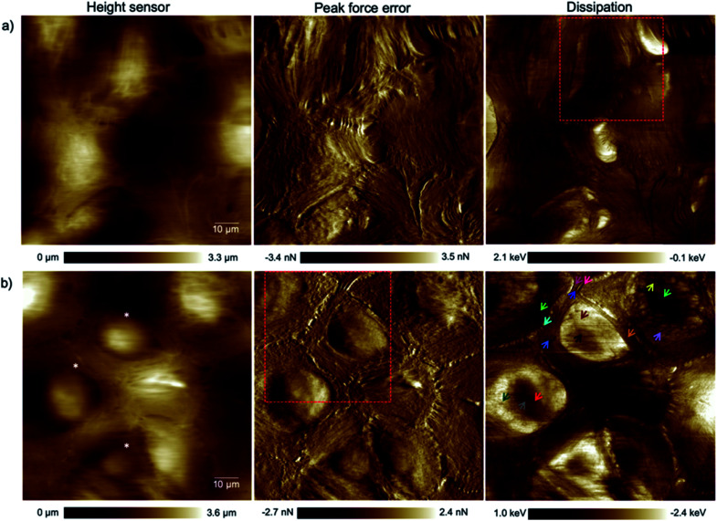
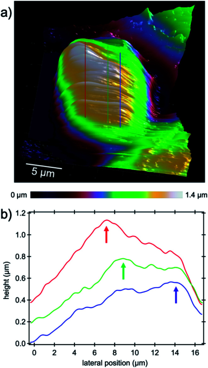
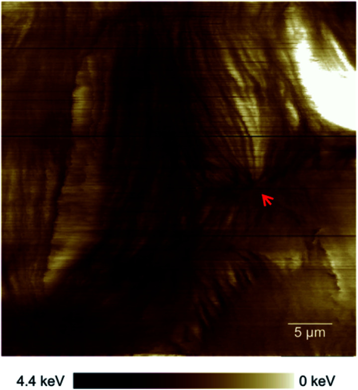
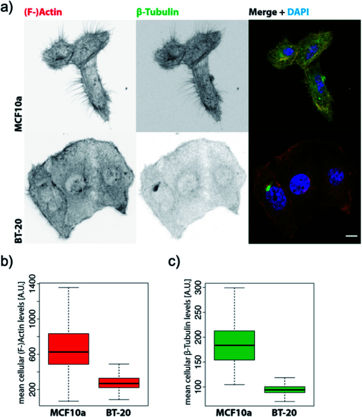
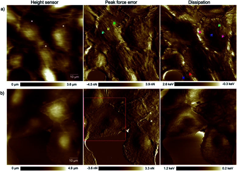
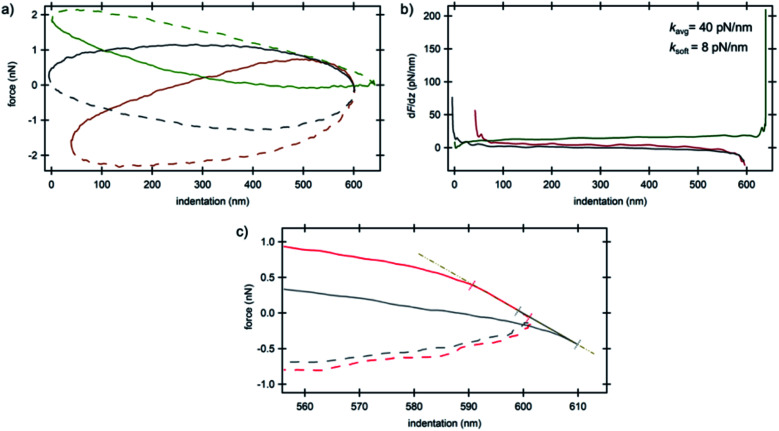
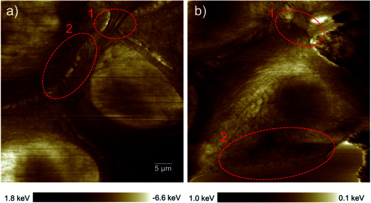
The transition of healthy epithelial cells to carcinoma is associated with an alteration in the structure and organization of the cytoskeleton of the cells. A comparison of the mechanical properties of cancerous and healthy cells indicated a higher deformability of the cancer cells based on averaging the mechanical properties of single cells. However, the exact reason for softening of the cancerous cells compared to their counterparts remains unclear. Here, we focused on nanomechanical spectroscopy of healthy and cancerous ductal epithelial-type breast cells by means of atomic force microscopy with high lateral and depth precision. As a result, based on atomic force microscopy measurements formation of significantly fewer microtubules in cancerous cells which was observed in our study is most likely one of the main causes for the overall change in mechanical properties without any phenotypic shift. Strikingly, in a confluent layer of invasive ductal carcinoma cells, we observed the formation of cell-cell junctions that have the potential for signal transduction among neighboring cells such as desmosomes and adherens junctions. This increases the possibility of cancerous cell collaboration in malignancy, infiltration or metastasis phenomena.

摘要

健康上皮细胞向癌细胞的转变与细胞细胞骨架结构和组织的改变有关。通过对癌细胞和健康细胞力学特性的比较,基于对单个细胞力学特性的平均,表明癌细胞具有更高的可变形性。然而,与健康细胞相比,癌细胞变软的确切原因仍不清楚。在此,我们通过具有高横向和深度精度的原子力显微镜,聚焦于健康和癌性导管上皮型乳腺细胞的纳米力学光谱分析。结果,基于原子力显微镜测量,我们研究中观察到癌细胞中微管形成显著减少,这很可能是机械性能整体变化而无任何表型转变的主要原因之一。引人注目的是,在浸润性导管癌细胞的汇合层中,我们观察到形成了细胞间连接,这些连接有可能在相邻细胞之间进行信号转导,如桥粒和黏着连接。这增加了癌细胞在恶性、浸润或转移现象中协作的可能性。

https://cdn.ncbi.nlm.nih.gov/pmc/blobs/4d7c/9418382/b7656ac428b2/c9na00021f-f7.jpg
https://cdn.ncbi.nlm.nih.gov/pmc/blobs/4d7c/9418382/f82b704ed105/c9na00021f-f1.jpg
https://cdn.ncbi.nlm.nih.gov/pmc/blobs/4d7c/9418382/14b8caccc582/c9na00021f-f2.jpg
https://cdn.ncbi.nlm.nih.gov/pmc/blobs/4d7c/9418382/2e56bee64dd4/c9na00021f-f3.jpg
https://cdn.ncbi.nlm.nih.gov/pmc/blobs/4d7c/9418382/40b268f3ed89/c9na00021f-f4.jpg
https://cdn.ncbi.nlm.nih.gov/pmc/blobs/4d7c/9418382/ae0ca08fea26/c9na00021f-f5.jpg
https://cdn.ncbi.nlm.nih.gov/pmc/blobs/4d7c/9418382/1656de1882dd/c9na00021f-f6.jpg
https://cdn.ncbi.nlm.nih.gov/pmc/blobs/4d7c/9418382/b7656ac428b2/c9na00021f-f7.jpg

相似文献

1
Structural analysis of healthy and cancerous epithelial-type breast cells by nanomechanical spectroscopy allows us to obtain peculiarities of the skeleton and junctions.通过纳米力学光谱对健康和癌性上皮型乳腺细胞进行结构分析,使我们能够获得细胞骨架和连接的特性。
Nanoscale Adv. 2019 Oct 25;1(12):4853-4862. doi: 10.1039/c9na00021f. eCollection 2019 Dec 3.
2
Viscoelastic properties of normal and cancerous human breast cells are affected differently by contact to adjacent cells.正常和癌变的人类乳腺细胞的黏弹性特性受到与相邻细胞接触的不同影响。
Acta Biomater. 2017 Jun;55:239-248. doi: 10.1016/j.actbio.2017.04.006. Epub 2017 Apr 7.
3
Atomic force microscope-based single cell force spectroscopy of breast cancer cell lines: an approach for evaluating cellular invasion.基于原子力显微镜的乳腺癌细胞系单细胞力谱分析:一种评估细胞侵袭的方法
J Biomech. 2014 Oct 17;47(13):3373-9. doi: 10.1016/j.jbiomech.2014.08.002. Epub 2014 Aug 12.
4
Nanomechanical sub-surface mapping of living biological cells by force microscopy.利用力显微镜对活生物细胞进行亚表面纳米力学测绘。
Nanoscale. 2019 Jul 21;11(27):13089-13097. doi: 10.1039/c9nr03497h. Epub 2019 Jul 3.
5
Formation of adherens junctions leads to the emergence of a tissue-level tension in epithelial monolayers.黏着连接的形成导致上皮单层中出现组织水平的张力。
J Cell Sci. 2014 Jun 1;127(Pt 11):2507-17. doi: 10.1242/jcs.142349. Epub 2014 Mar 21.
6
The softening of human bladder cancer cells happens at an early stage of the malignancy process.人类膀胱癌细胞的软化发生在恶性肿瘤过程的早期。
Beilstein J Nanotechnol. 2014 Apr 10;5:447-57. doi: 10.3762/bjnano.5.52. eCollection 2014.
7
Importance of integrity of cell-cell junctions for the mechanics of confluent MDCK II cells.细胞-细胞连接完整性对 MDCK II 细胞汇合力学的重要性。
Sci Rep. 2018 Sep 20;8(1):14117. doi: 10.1038/s41598-018-32421-2.
8
Effect of Actin Organization on the Stiffness of Living Breast Cancer Cells Revealed by Peak-Force Modulation Atomic Force Microscopy.峰值力调制原子力显微镜揭示了肌动蛋白组织对活体乳腺癌细胞刚性的影响。
ACS Nano. 2016 Mar 22;10(3):3365-74. doi: 10.1021/acsnano.5b07162. Epub 2016 Feb 25.
9
The cyto-linker and scaffolding protein "plectin" mis-localization leads to softening of cancer cells.细胞连接蛋白和支架蛋白“网蛋白”的定位错误导致癌细胞变软。
Nanoscale. 2023 Sep 21;15(36):15008-15026. doi: 10.1039/d3nr02226a.
10
Cell-cell junctions as sensors and transducers of mechanical forces.细胞-细胞连接作为机械力的感受器和转换器。
Biochim Biophys Acta Biomembr. 2020 Sep 1;1862(9):183316. doi: 10.1016/j.bbamem.2020.183316. Epub 2020 Apr 28.

引用本文的文献

1
Mechanical Properties and Nanomotion of BT-20 and ZR-75 Breast Cancer Cells Studied by Atomic Force Microscopy and Optical Nanomotion Detection Method.原子力显微镜和光学纳米运动检测法研究 BT-20 和 ZR-75 乳腺癌细胞的力学性能和纳米运动。
Cells. 2023 Sep 26;12(19):2362. doi: 10.3390/cells12192362.

本文引用的文献

1
Nanomechanical sub-surface mapping of living biological cells by force microscopy.利用力显微镜对活生物细胞进行亚表面纳米力学测绘。
Nanoscale. 2019 Jul 21;11(27):13089-13097. doi: 10.1039/c9nr03497h. Epub 2019 Jul 3.
2
Real Microgravity Influences the Cytoskeleton and Focal Adhesions in Human Breast Cancer Cells.真正的微重力会影响人乳腺癌细胞的细胞骨架和焦点黏附。
Int J Mol Sci. 2019 Jun 28;20(13):3156. doi: 10.3390/ijms20133156.
3
Phenotypic and molecular dissection of metaplastic breast cancer and the prognostic implications.
探讨化生性乳腺癌的表型和分子特征及其预后意义。
J Pathol. 2019 Feb;247(2):214-227. doi: 10.1002/path.5184. Epub 2018 Dec 20.
4
Determination of the Elastic Moduli of a Single Cell Cultured on a Rigid Support by Force Microscopy.通过力显微镜测定刚性基底上培养的单细胞的弹性模量。
Biophys J. 2018 Jun 19;114(12):2923-2932. doi: 10.1016/j.bpj.2018.05.012.
5
Ribosome biogenesis in cancer: new players and therapeutic avenues.癌症中的核糖体生物发生:新的参与者和治疗途径。
Nat Rev Cancer. 2018 Jan;18(1):51-63. doi: 10.1038/nrc.2017.104. Epub 2017 Dec 1.
6
Breast Cancer Cell Line Classification and Its Relevance with Breast Tumor Subtyping.乳腺癌细胞系分类及其与乳腺肿瘤亚型的相关性。
J Cancer. 2017 Sep 12;8(16):3131-3141. doi: 10.7150/jca.18457. eCollection 2017.
7
Matrix stiffness induces epithelial-mesenchymal transition and promotes chemoresistance in pancreatic cancer cells.基质硬度诱导胰腺癌细胞发生上皮-间质转化并促进其化疗耐药性。
Oncogenesis. 2017 Jul 3;6(7):e352. doi: 10.1038/oncsis.2017.54.
8
Synergistic IL-6 and IL-8 paracrine signalling pathway infers a strategy to inhibit tumour cell migration.协同的 IL-6 和 IL-8 旁分泌信号通路提示了抑制肿瘤细胞迁移的策略。
Nat Commun. 2017 May 26;8:15584. doi: 10.1038/ncomms15584.
9
L1 retrotransposition is activated by Ten-eleven-translocation protein 1 and repressed by methyl-CpG binding proteins.L1 retrotransposition 被 Ten-eleven-translocation protein 1 激活,并被甲基-CpG 结合蛋白抑制。
Nucleus. 2017 Sep 3;8(5):548-562. doi: 10.1080/19491034.2017.1330238. Epub 2017 May 19.
10
Biomechanics of subcellular structures by non-invasive Brillouin microscopy.通过非侵入性布里渊显微镜研究亚细胞结构的生物力学
Sci Rep. 2016 Nov 15;6:37217. doi: 10.1038/srep37217.