• 文献检索
  • 文档翻译
  • 深度研究
  • 学术资讯
  • Suppr Zotero 插件Zotero 插件
  • 邀请有礼
  • 套餐&价格
  • 历史记录
应用&插件
Suppr Zotero 插件Zotero 插件浏览器插件Mac 客户端Windows 客户端微信小程序
定价
高级版会员购买积分包购买API积分包
服务
文献检索文档翻译深度研究API 文档MCP 服务
关于我们
关于 Suppr公司介绍联系我们用户协议隐私条款
关注我们

Suppr 超能文献

核心技术专利:CN118964589B侵权必究
粤ICP备2023148730 号-1Suppr @ 2026

文献检索

告别复杂PubMed语法,用中文像聊天一样搜索,搜遍4000万医学文献。AI智能推荐,让科研检索更轻松。

立即免费搜索

文件翻译

保留排版,准确专业,支持PDF/Word/PPT等文件格式,支持 12+语言互译。

免费翻译文档

深度研究

AI帮你快速写综述,25分钟生成高质量综述,智能提取关键信息,辅助科研写作。

立即免费体验

从病理学到 MRI 再到病理学:多发性硬化病变的临床相关生物标志物。

From pathology to MRI and back: Clinically relevant biomarkers of multiple sclerosis lesions.

机构信息

Translational Neuroradiology Section, National Institute of Neurological Disorders and Stroke (NINDS), National Institutes of Health (NIH), Bethesda, MD, USA; Department of Neurology, Tel Aviv Sourasky Medical Center, Tel Aviv-Yaffo, Israel.

Translational Neuroradiology Section, National Institute of Neurological Disorders and Stroke (NINDS), National Institutes of Health (NIH), Bethesda, MD, USA; Cedars-Sinai Medical Center, Los Angeles, CA, USA.

出版信息

Neuroimage Clin. 2022;36:103194. doi: 10.1016/j.nicl.2022.103194. Epub 2022 Sep 14.

DOI:10.1016/j.nicl.2022.103194
PMID:36170753
原文链接:https://pmc.ncbi.nlm.nih.gov/articles/PMC9668624/
Abstract

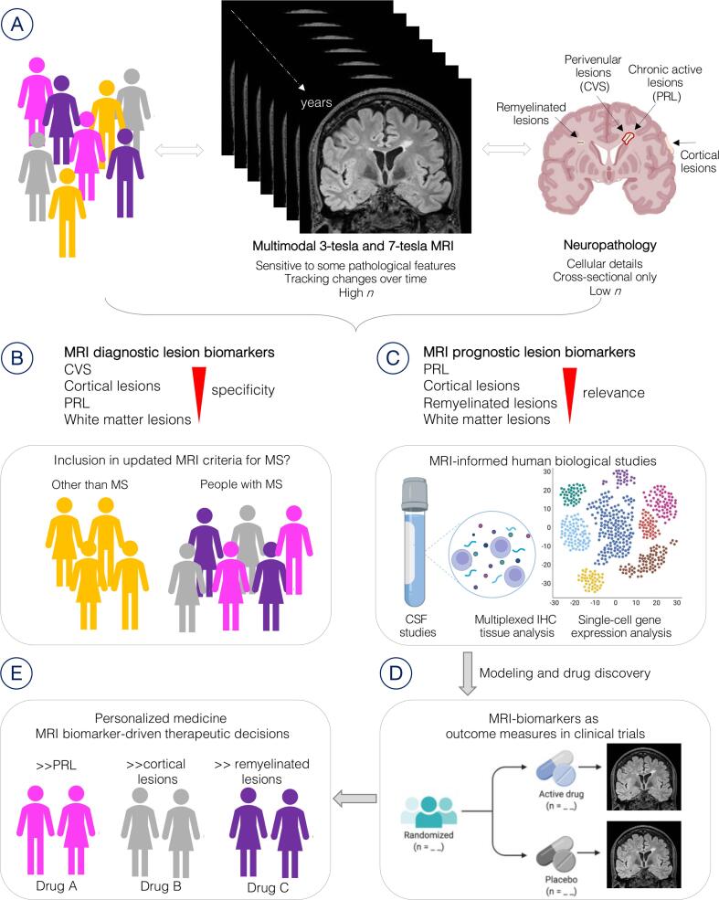
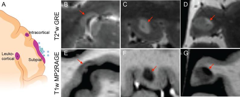
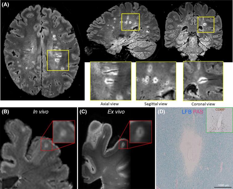
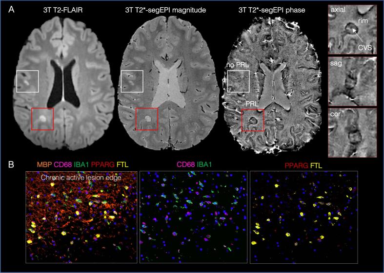
Focal lesions in both white and gray matter are characteristic of multiple sclerosis (MS). Histopathological studies have helped define the main underlying pathological processes involved in lesion formation and evolution, serving as a gold standard for many years. However, histopathology suffers from an intrinsic bias resulting from over-reliance on tissue samples from late stages of the disease or atypical cases and is inadequate for routine patient assessment. Pathological-radiological correlative studies have established advanced MRI's sensitivity to several relevant MS-pathological substrates and its practicality for assessing dynamic changes and following lesions over time. This review focuses on novel imaging techniques that serve as biomarkers of critical pathological substrates of MS lesions: the central vein, chronic inflammation, remyelination and repair, and cortical lesions. For each pathological process, we address the correlative value of MRI to MS pathology, its contribution in elucidating MS pathology in vivo, and the clinical utility of the imaging biomarker.

摘要

大脑白质和灰质中的局灶性病变是多发性硬化症(MS)的特征。组织病理学研究有助于确定病变形成和演变所涉及的主要潜在病理过程,多年来一直是金标准。然而,组织病理学存在内在偏差,过度依赖疾病晚期或非典型病例的组织样本,因此不足以进行常规患者评估。病理-放射学相关性研究已经确定了高级 MRI 对几种相关的 MS 病理基质的敏感性及其用于评估动态变化和随时间推移跟踪病变的实用性。这篇综述重点介绍了作为 MS 病变关键病理基质的生物标志物的新型成像技术:中央静脉、慢性炎症、髓鞘再生和修复以及皮质病变。对于每个病理过程,我们将探讨 MRI 对 MS 病理的相关性、其在体内阐明 MS 病理方面的贡献以及成像生物标志物的临床实用性。

https://cdn.ncbi.nlm.nih.gov/pmc/blobs/0d88/9668624/270e945c924f/gr5.jpg
https://cdn.ncbi.nlm.nih.gov/pmc/blobs/0d88/9668624/28f6b248688d/gr1.jpg
https://cdn.ncbi.nlm.nih.gov/pmc/blobs/0d88/9668624/5e3ecc316d77/gr2.jpg
https://cdn.ncbi.nlm.nih.gov/pmc/blobs/0d88/9668624/25babe2e0b82/gr3.jpg
https://cdn.ncbi.nlm.nih.gov/pmc/blobs/0d88/9668624/cc176eea7bff/gr4.jpg
https://cdn.ncbi.nlm.nih.gov/pmc/blobs/0d88/9668624/270e945c924f/gr5.jpg

相似文献

1
From pathology to MRI and back: Clinically relevant biomarkers of multiple sclerosis lesions.从病理学到 MRI 再到病理学:多发性硬化病变的临床相关生物标志物。
Neuroimage Clin. 2022;36:103194. doi: 10.1016/j.nicl.2022.103194. Epub 2022 Sep 14.
2
Association of Chronic Active Multiple Sclerosis Lesions With Disability In Vivo.慢性活动性多发性硬化症病灶与体内残疾的关联。
JAMA Neurol. 2019 Dec 1;76(12):1474-1483. doi: 10.1001/jamaneurol.2019.2399.
3
Association between pathological and MRI findings in multiple sclerosis.多发性硬化的病理与 MRI 表现的相关性。
Lancet Neurol. 2019 Feb;18(2):198-210. doi: 10.1016/S1474-4422(18)30451-4.
4
Imaging the multiple sclerosis lesion: insights into pathogenesis, progression and repair.多发性硬化症病灶的影像学研究:对发病机制、进展和修复的深入了解。
Curr Opin Neurol. 2019 Jun;32(3):338-345. doi: 10.1097/WCO.0000000000000698.
5
Central Vein Sign and Paramagnetic Rim Lesions: Susceptibility Changes in Brain Tissues and Their Implications for the Study of Multiple Sclerosis Pathology.中央静脉征与顺磁性边缘病变:脑组织的磁化率变化及其对多发性硬化症病理学研究的意义
Diagnostics (Basel). 2024 Jun 27;14(13):1362. doi: 10.3390/diagnostics14131362.
6
Imaging in multiple sclerosis: A new spin on lesions.多发性硬化症的影像学:病灶的新视角。
J Med Imaging Radiat Oncol. 2016 Oct;60(5):577-586. doi: 10.1111/1754-9485.12498. Epub 2016 Jul 27.
7
Association between pathological and MRI findings in multiple sclerosis.多发性硬化的病理与 MRI 表现的相关性。
Lancet Neurol. 2012 Apr;11(4):349-60. doi: 10.1016/S1474-4422(12)70003-0. Epub 2012 Mar 19.
8
Longitudinal evaluation of demyelinated lesions in a multiple sclerosis model using ultrashort echo time magnetization transfer (UTE-MT) imaging.使用超短回波时间磁化传递(UTE-MT)成像对多发性硬化模型中的脱髓鞘病变进行纵向评估。
Neuroimage. 2020 Mar;208:116415. doi: 10.1016/j.neuroimage.2019.116415. Epub 2019 Dec 4.
9
Evaluation of the Central Vein Sign as a Diagnostic Imaging Biomarker in Multiple Sclerosis.中央静脉征作为多发性硬化症诊断影像学生物标志物的评估。
JAMA Neurol. 2019 Dec 1;76(12):1446-1456. doi: 10.1001/jamaneurol.2019.2478.
10
Predicting PET-derived myelin content from multisequence MRI for individual longitudinal analysis in multiple sclerosis.从多序列 MRI 预测 PET 衍生的髓鞘含量,用于多发性硬化症的个体纵向分析。
Neuroimage. 2020 Dec;223:117308. doi: 10.1016/j.neuroimage.2020.117308. Epub 2020 Sep 2.

引用本文的文献

1
Validation of retroactively derived T1 relaxation values from 3D T1-weighted images with clinical and MRI measures of disability in multiple sclerosis.通过临床和MRI残疾测量指标对从三维T1加权图像追溯得出的T1弛豫值进行验证:多发性硬化症研究
PLoS One. 2025 May 19;20(5):e0323898. doi: 10.1371/journal.pone.0323898. eCollection 2025.
2
Current challenges in secondary progressive multiple sclerosis: diagnosis, activity detection and treatment.继发进展型多发性硬化的当前挑战:诊断、活动检测与治疗
Front Immunol. 2025 Mar 21;16:1543649. doi: 10.3389/fimmu.2025.1543649. eCollection 2025.
3
Towards a biological view of multiple sclerosis from early subtle to clinical progression: an expert opinion.
从早期细微变化到临床进展,构建多发性硬化症的生物学视角:专家意见
J Neurol. 2025 Feb 1;272(2):179. doi: 10.1007/s00415-025-12917-4.
4
Perceptual super-resolution in multiple sclerosis MRI.多发性硬化症磁共振成像中的感知超分辨率
Front Neurosci. 2024 Oct 22;18:1473132. doi: 10.3389/fnins.2024.1473132. eCollection 2024.
5
Predicting multiple sclerosis disease progression and outcomes with machine learning and MRI-based biomarkers: a review.利用机器学习和基于MRI的生物标志物预测多发性硬化症的疾病进展和预后:综述
J Neurol. 2024 Oct;271(10):6543-6572. doi: 10.1007/s00415-024-12651-3. Epub 2024 Sep 12.
6
Decoding Gray Matter Involvement in Multiple Sclerosis via Imaging.通过成像技术解码多发性硬化症中灰质的受累情况。
Neuroimaging Clin N Am. 2024 Aug;34(3):453-468. doi: 10.1016/j.nic.2024.03.007. Epub 2024 May 19.
7
Advanced MRI Techniques: Diagnosis and Follow-Up of Multiple Sclerosis.先进的磁共振成像技术:多发性硬化症的诊断与随访
Diagnostics (Basel). 2024 May 28;14(11):1120. doi: 10.3390/diagnostics14111120.
8
Neurodegeneration and demyelination in multiple sclerosis.多发性硬化中的神经退行性变和脱髓鞘。
Neuron. 2024 Oct 9;112(19):3231-3251. doi: 10.1016/j.neuron.2024.05.025. Epub 2024 Jun 17.
9
Neuroimaging assessment of facility-bound severely-affected MS reveals the critical role of cortical gray matter pathology: results from the CASA-MS case-controlled study.对机构内严重受影响的多发性硬化症患者的神经影像学评估揭示了皮质灰质病变的关键作用:来自CASA-MS病例对照研究的结果
J Neurol. 2024 Aug;271(8):4949-4962. doi: 10.1007/s00415-024-12420-2. Epub 2024 May 17.
10
Emerging imaging markers in radiologically isolated syndrome: implications for earlier treatment initiation.影像学孤立综合征中的新兴标志物:对早期治疗启动的影响。
Neurol Sci. 2024 Jul;45(7):3061-3068. doi: 10.1007/s10072-024-07402-1. Epub 2024 Feb 19.