• 文献检索
  • 文档翻译
  • 深度研究
  • 学术资讯
  • Suppr Zotero 插件Zotero 插件
  • 邀请有礼
  • 套餐&价格
  • 历史记录
应用&插件
Suppr Zotero 插件Zotero 插件浏览器插件Mac 客户端Windows 客户端微信小程序
定价
高级版会员购买积分包购买API积分包
服务
文献检索文档翻译深度研究API 文档MCP 服务
关于我们
关于 Suppr公司介绍联系我们用户协议隐私条款
关注我们

Suppr 超能文献

核心技术专利:CN118964589B侵权必究
粤ICP备2023148730 号-1Suppr @ 2026

文献检索

告别复杂PubMed语法,用中文像聊天一样搜索,搜遍4000万医学文献。AI智能推荐,让科研检索更轻松。

立即免费搜索

文件翻译

保留排版,准确专业,支持PDF/Word/PPT等文件格式,支持 12+语言互译。

免费翻译文档

深度研究

AI帮你快速写综述,25分钟生成高质量综述,智能提取关键信息,辅助科研写作。

立即免费体验

MRI 可见血管周围空间与痴呆发病风险:弗雷明汉心脏研究。

MRI-Visible Perivascular Spaces and Risk of Incident Dementia: The Framingham Heart Study.

机构信息

From the Department of Neurology (J.R.R., H.J.A.), Boston University School of Medicine, MA; Department of Biostatistics (A.P. S.D.), Boston University School of Public Health, MA; Department of Neurology (C.S.D.), University of California at Davis, CA; The Glenn Biggs Institute for Alzheimer's and Neurodegenerative Diseases (S.S.), University of Texas Health Sciences Center, San Antonio, TX; and NHLBI's Framingham Heart Study (J.R.R., A.P., H.J.A., S.D., S.S.), MA.

出版信息

Neurology. 2022 Dec 5;99(23):e2561-e2571. doi: 10.1212/WNL.0000000000201293.

DOI:10.1212/WNL.0000000000201293
PMID:36175148
原文链接:https://pmc.ncbi.nlm.nih.gov/articles/PMC9754649/
Abstract

BACKGROUND AND OBJECTIVES

Perivascular spaces (PVS) visible on MRI scans may represent key aspects in the pathophysiology of stroke and dementia, including cerebral small vessel disease and glymphatic dysfunction. This study aimed to determine the association between MRI-visible PVS burden and the risk of incident dementia.

METHODS

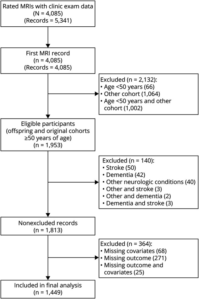
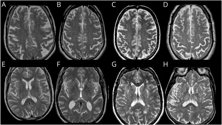
This study included community-dwelling Framingham Heart Study Original and Offspring cohort participants with available brain MRI-PVS ratings, free of stroke and dementia. Multivariable Cox proportional hazard regression was used to obtain hazard ratios (HRs) and 95% CIs of the association between MRI-visible PVS and incident dementia. PVS were rated using validated methods in the basal ganglia (BG) and centrum semiovale (CSO). The outcomes included all-cause dementia, Alzheimer dementia (AD), and vascular dementia (VaD).

RESULTS

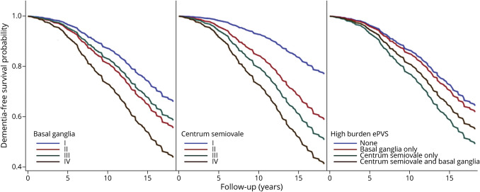
One thousand four hundred forty-nine participants 50 years or older (46% male) were included. Over a median follow-up period of 8.3 years, the incidence of all-cause dementia, AD, and VaD was 15.8%, 12.5%, and 2.5%, respectively. In models that adjusted for vascular risk factors and cardiovascular disease, the hazard for dementia increased steadily as PVS burden increased, rising 2-fold for those with grade II PVS (HR 2.44, 95% CI 1.51-3.93) to 5-fold in participants with grade IV (HR 5.05, 95% CI 2.75-9.26) compared with grade I PVS in CSO. In the BG, hazards increased 1.6-fold (HR 1.62, 95% CI 1.15-2.27) for grade II to 2.6-fold (HR 2.67, 95% CI 1.04-6.88) for grade IV compared with grade I PVS. The association remained significant for CSO but not for BG, after adjustment for white matter hyperintensity volume (WMHV), covert infarcts, and total brain volume. Similar findings were observed for AD, but VaD, limited by a small number of events, was not statistically significant.

DISCUSSION

Higher burden of PVS in CSO was associated with increased risk of developing dementia, independent of vascular risk factors, total brain volume, WMHVs, and covert infarcts. This finding supports a role for PVS as a subclinical MRI marker to identify individuals in subclinical stages at high risk of developing dementia who may benefit from early intervention.

摘要

背景与目的

磁共振成像(MRI)扫描中可见的血管周围间隙(PVS)可能代表中风和痴呆病理生理学中的关键方面,包括脑小血管疾病和糖质淋系统功能障碍。本研究旨在确定 MRI 可见 PVS 负担与痴呆症发病风险之间的关联。

方法

本研究纳入了Framingham 心脏研究原队列和后代队列中居住在社区的参与者,这些参与者具有可用的脑 MRI-PVS 评分,且无中风和痴呆。采用多变量 Cox 比例风险回归分析评估 MRI 可见 PVS 与新发痴呆之间的关联。使用基底节(BG)和半卵圆中心(CSO)的验证方法对 PVS 进行评分。结局包括全因痴呆、阿尔茨海默病痴呆(AD)和血管性痴呆(VaD)。

结果

本研究共纳入了 1449 名 50 岁及以上(46%为男性)的参与者。在中位随访 8.3 年期间,全因痴呆、AD 和 VaD 的发生率分别为 15.8%、12.5%和 2.5%。在调整血管危险因素和心血管疾病的模型中,随着 PVS 负担的增加,痴呆的风险呈稳步上升趋势,CSO 中 PVS 分级 II 级(HR 2.44,95%CI 1.51-3.93)和 IV 级(HR 5.05,95%CI 2.75-9.26)的风险分别是 PVS 分级 I 级的 2 倍和 5 倍。在 BG 中,PVS 分级 II 级(HR 1.62,95%CI 1.15-2.27)和 IV 级(HR 2.67,95%CI 1.04-6.88)的风险分别是 PVS 分级 I 级的 1.6 倍和 2.6 倍。在调整脑白质高信号容积(WMHV)、隐匿性梗死和总脑容量后,CSO 中这种关联仍然显著,但在 BG 中则不显著。类似的发现也见于 AD,但由于事件数量有限,VaD 无统计学意义。

讨论

CSO 中 PVS 负担的增加与痴呆风险的增加相关,这独立于血管危险因素、总脑容量、WMHV 和隐匿性梗死。这一发现支持 PVS 作为一种亚临床 MRI 标志物的作用,可用于识别处于亚临床阶段、痴呆风险较高的个体,这些个体可能受益于早期干预。

https://cdn.ncbi.nlm.nih.gov/pmc/blobs/53cf/9754649/13aa62dd0816/WNL-2022-201192f3.jpg
https://cdn.ncbi.nlm.nih.gov/pmc/blobs/53cf/9754649/58b4edbd3a80/WNL-2022-201192f1.jpg
https://cdn.ncbi.nlm.nih.gov/pmc/blobs/53cf/9754649/02605c7acaea/WNL-2022-201192f2.jpg
https://cdn.ncbi.nlm.nih.gov/pmc/blobs/53cf/9754649/13aa62dd0816/WNL-2022-201192f3.jpg
https://cdn.ncbi.nlm.nih.gov/pmc/blobs/53cf/9754649/58b4edbd3a80/WNL-2022-201192f1.jpg
https://cdn.ncbi.nlm.nih.gov/pmc/blobs/53cf/9754649/02605c7acaea/WNL-2022-201192f2.jpg
https://cdn.ncbi.nlm.nih.gov/pmc/blobs/53cf/9754649/13aa62dd0816/WNL-2022-201192f3.jpg

相似文献

1
MRI-Visible Perivascular Spaces and Risk of Incident Dementia: The Framingham Heart Study.MRI 可见血管周围空间与痴呆发病风险:弗雷明汉心脏研究。
Neurology. 2022 Dec 5;99(23):e2561-e2571. doi: 10.1212/WNL.0000000000201293.
2
Association of MRI Visible Perivascular Spaces and Neurofilament Light Chain: The Framingham Heart Study.MRI 可见血管周围空间与神经丝轻链的关联:弗雷明汉心脏研究。
J Alzheimers Dis. 2023;95(3):1133-1145. doi: 10.3233/JAD-221260.
3
MRI Visible Perivascular Spaces and the Risk of Incident Mild Cognitive Impairment in a Community Sample.MRI 可见血管周围空间与社区样本中轻度认知障碍事件风险的关系。
J Alzheimers Dis. 2023;96(1):103-112. doi: 10.3233/JAD-230445.
4
Inflammatory biomarkers and MRI visible perivascular spaces: The Framingham Heart Study.炎症生物标志物与 MRI 可见血管周围间隙:弗雷明汉心脏研究。
Neurobiol Aging. 2023 Jul;127:12-22. doi: 10.1016/j.neurobiolaging.2023.03.001. Epub 2023 Mar 6.
5
Relation of MRI-Visible Perivascular Spaces and Other MRI Markers of Cerebral Small Vessel Disease.脑小血管病的MRI可见血管周围间隙与其他MRI标志物的关系。
Brain Sci. 2023 Sep 14;13(9):1323. doi: 10.3390/brainsci13091323.
6
Associations of MRI-visible perivascular spaces with longitudinal cognitive decline across the Alzheimer's disease spectrum.MRI 可见血管周围空间与阿尔茨海默病谱中纵向认知衰退的关联。
Alzheimers Res Ther. 2022 Dec 13;14(1):185. doi: 10.1186/s13195-022-01136-y.
7
Topography and Determinants of Magnetic Resonance Imaging (MRI)-Visible Perivascular Spaces in a Large Memory Clinic Cohort.磁共振成像(MRI)可见血管周围空间的地形学及其决定因素:一项大型记忆门诊队列研究。
J Am Heart Assoc. 2017 Sep 22;6(9):e006279. doi: 10.1161/JAHA.117.006279.
8
Association of Dilated Perivascular Spaces With Cognitive Decline and Incident Dementia.血管周围间隙扩张与认知能力下降和痴呆发病的关系。
Neurology. 2021 Mar 16;96(11):e1501-e1511. doi: 10.1212/WNL.0000000000011537. Epub 2021 Jan 27.
9
Clinical Significance of Magnetic Resonance Imaging Markers of Vascular Brain Injury: A Systematic Review and Meta-analysis.磁共振血管损伤标志物的临床意义:系统评价和荟萃分析。
JAMA Neurol. 2019 Jan 1;76(1):81-94. doi: 10.1001/jamaneurol.2018.3122.
10
Associations Between Vascular Risk Factors and Perivascular Spaces in Adults with Intact Cognition, Mild Cognitive Impairment, and Dementia.血管危险因素与认知正常、轻度认知障碍和痴呆成人的血管周围间隙的关系。
J Alzheimers Dis. 2022;89(2):437-448. doi: 10.3233/JAD-215585.

引用本文的文献

1
Multimarker Cerebral Small Vessel Disease Score and Risk of Incident Dementia in the Framingham Heart Study.弗雷明汉心脏研究中的多标志物脑小血管疾病评分与新发痴呆风险
Neurology. 2025 Oct 7;105(7):e214113. doi: 10.1212/WNL.0000000000214113. Epub 2025 Sep 15.
2
Relationships between measures of neurovascular integrity and fluid transport in aging: a multi-modal neuroimaging study.衰老过程中神经血管完整性指标与液体运输之间的关系:一项多模态神经影像学研究。
Fluids Barriers CNS. 2025 Jun 16;22(1):59. doi: 10.1186/s12987-025-00664-7.
3
Association of Serum Neurofilament Light and Glial Fibrillary Acidic Protein with Cerebral Small Vessel Disease in Nondemented Middle-to-Old Aged Population.
血清神经丝轻链和胶质纤维酸性蛋白与非痴呆中老年人群脑小血管病的关联
Transl Stroke Res. 2025 May 31. doi: 10.1007/s12975-025-01359-9.
4
Cerebral small vessel disease among rural-dwelling Chinese older adults: prevalence, distribution, and associated factors.中国农村老年人群中的脑小血管病:患病率、分布及相关因素
Brain Commun. 2025 Apr 4;7(2):fcaf136. doi: 10.1093/braincomms/fcaf136. eCollection 2025.
5
Cerebrovascular burden and neurodegeneration linked to 15-year odor identification decline in older adults.脑血管负担和神经退行性变与老年人15年的气味识别能力下降有关。
Front Aging Neurosci. 2025 Mar 24;17:1539508. doi: 10.3389/fnagi.2025.1539508. eCollection 2025.
6
Imaging markers of cerebral small vessel disease are associated with Alzheimer's disease: a systematic review and meta-analysis.脑小血管病的影像学标志物与阿尔茨海默病相关:一项系统评价和荟萃分析。
Front Aging Neurosci. 2025 Jan 28;17:1498636. doi: 10.3389/fnagi.2025.1498636. eCollection 2025.
7
Relation of MRI visible perivascular spaces with global and regional brain structural connectivity measures: The Framingham Heart Study (FHS).磁共振成像可见的血管周围间隙与全脑和局部脑结构连接性测量指标的关系:弗雷明汉心脏研究(FHS)
Neurobiol Aging. 2025 Jun;150:1-8. doi: 10.1016/j.neurobiolaging.2025.02.007. Epub 2025 Feb 27.
8
Robust, fully-automated assessment of cerebral perivascular spaces and white matter lesions: a multicentre MRI longitudinal study of their evolution and association with risk of dementia and accelerated brain atrophy.脑周血管间隙和白质病变的稳健、全自动评估:一项关于其演变以及与痴呆风险和脑萎缩加速相关性的多中心MRI纵向研究
EBioMedicine. 2025 Jan;111:105523. doi: 10.1016/j.ebiom.2024.105523. Epub 2024 Dec 24.
9
Cerebrovascular markers of WMH and infarcts in ADNI: A historical perspective and future directions.ADNI中脑白质高信号和梗死灶的脑血管标志物:历史回顾与未来方向
Alzheimers Dement. 2024 Dec;20(12):8953-8968. doi: 10.1002/alz.14358. Epub 2024 Nov 13.
10
Perivascular space dysfunction in cerebral small vessel disease is related to neuroinflammation.脑小血管病中的血管周围间隙功能障碍与神经炎症有关。
Brain. 2025 May 13;148(5):1540-1550. doi: 10.1093/brain/awae357.