• 文献检索
  • 文档翻译
  • 深度研究
  • 学术资讯
  • Suppr Zotero 插件Zotero 插件
  • 邀请有礼
  • 套餐&价格
  • 历史记录
应用&插件
Suppr Zotero 插件Zotero 插件浏览器插件Mac 客户端Windows 客户端微信小程序
定价
高级版会员购买积分包购买API积分包
服务
文献检索文档翻译深度研究API 文档MCP 服务
关于我们
关于 Suppr公司介绍联系我们用户协议隐私条款
关注我们

Suppr 超能文献

核心技术专利:CN118964589B侵权必究
粤ICP备2023148730 号-1Suppr @ 2026

文献检索

告别复杂PubMed语法,用中文像聊天一样搜索,搜遍4000万医学文献。AI智能推荐,让科研检索更轻松。

立即免费搜索

文件翻译

保留排版,准确专业,支持PDF/Word/PPT等文件格式,支持 12+语言互译。

免费翻译文档

深度研究

AI帮你快速写综述,25分钟生成高质量综述,智能提取关键信息,辅助科研写作。

立即免费体验

脑小血管病的MRI可见血管周围间隙与其他MRI标志物的关系。

Relation of MRI-Visible Perivascular Spaces and Other MRI Markers of Cerebral Small Vessel Disease.

作者信息

Rodriguez Lara Frances, Toro Arturo Ruben, Pinheiro Adlin, Demissie Serkalem, Ekenze Oluchi, Martinez Oliver, Parva Pedram, Charidimou Andreas, Ghosh Saptaparni, DeCarli Charles, Seshadri Sudha, Habes Mohamad, Maillard Pauline, Romero Jose Rafael

机构信息

Chobanian & Avedisian School of Medicine, Boston University, Boston, MA 02118, USA.

Department of Biostatistics, School of Public Health, Boston University, Boston, MA 02118, USA.

出版信息

Brain Sci. 2023 Sep 14;13(9):1323. doi: 10.3390/brainsci13091323.

DOI:10.3390/brainsci13091323
PMID:37759924
原文链接:https://pmc.ncbi.nlm.nih.gov/articles/PMC10527297/
Abstract

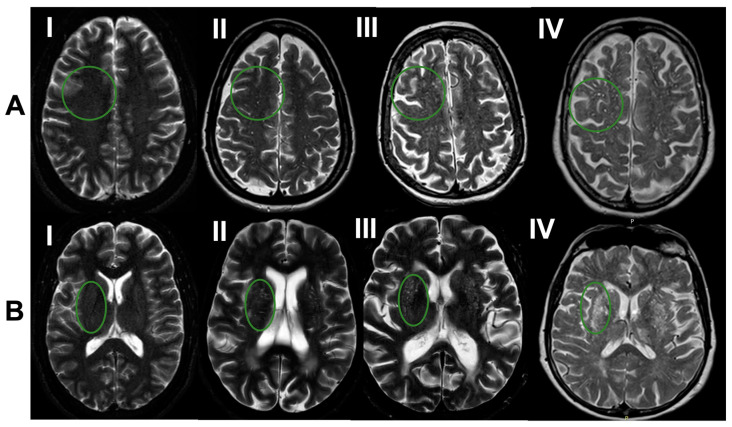
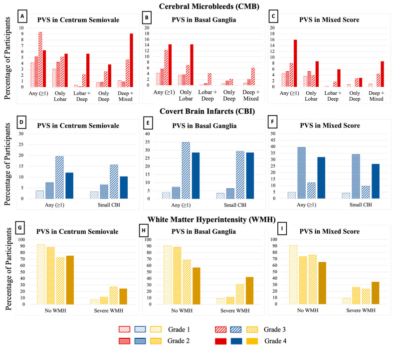
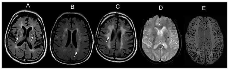
Perivascular spaces (PVS) visible on brain MRI signal cerebral small vessel disease (CSVD). The coexistence of PVS with other CSVD manifestations likely increases the risk of adverse neurological outcomes. We related PVS to other CSVD manifestations and brain volumes that are markers of vascular brain injury and neurodegeneration. Framingham Heart Study (FHS) participants with CSVD ratings on brain MRI were included. PVS were rated in the basal ganglia (BG) and centrum semiovale (CSO) into grades I-IV and a category reflecting high burden in single or mixed CSO-BG regions. We related PVS to covert brain infarcts (CBI), white matter hyperintensities (WMH), cerebral microbleeds (CMB), total brain, hippocampal, and cortical gray matter volumes using adjusted multivariable regression analyses. In 2454 participants (mean age 54 ± 12 years), we observed that higher PVS burden in both BG and CSO was related to CMB in lobar and deep brain regions and increased WMH. Greater CSO PVS burden was associated with decreased total cortical gray volumes. PVS are associated with ischemic markers of CSVD and neurodegeneration markers. Further studies should elucidate the causality between PVS and other CSVD manifestations.

摘要

脑磁共振成像(MRI)上可见的血管周围间隙(PVS)提示脑小血管病(CSVD)。PVS与其他CSVD表现共存可能会增加不良神经结局的风险。我们将PVS与其他CSVD表现以及作为血管性脑损伤和神经退行性变标志物的脑容量进行了关联研究。纳入了在脑MRI上有CSVD评分的弗雷明汉心脏研究(FHS)参与者。对基底节(BG)和半卵圆中心(CSO)的PVS进行分级,分为I-IV级以及反映单个或混合CSO-BG区域高负荷的类别。我们使用校正后的多变量回归分析将PVS与隐匿性脑梗死(CBI)、白质高信号(WMH)、脑微出血(CMB)、全脑、海马和皮质灰质体积进行关联。在2454名参与者(平均年龄54±12岁)中,我们观察到BG和CSO中较高的PVS负荷与脑叶和深部脑区的CMB以及增加的WMH相关。CSO中更大的PVS负荷与总皮质灰质体积减少相关。PVS与CSVD的缺血标志物和神经退行性变标志物相关。进一步的研究应阐明PVS与其他CSVD表现之间的因果关系。

https://cdn.ncbi.nlm.nih.gov/pmc/blobs/21f1/10527297/114e7c505683/brainsci-13-01323-g003.jpg
https://cdn.ncbi.nlm.nih.gov/pmc/blobs/21f1/10527297/6469cdf16c24/brainsci-13-01323-g001.jpg
https://cdn.ncbi.nlm.nih.gov/pmc/blobs/21f1/10527297/133e914cff75/brainsci-13-01323-g002.jpg
https://cdn.ncbi.nlm.nih.gov/pmc/blobs/21f1/10527297/114e7c505683/brainsci-13-01323-g003.jpg
https://cdn.ncbi.nlm.nih.gov/pmc/blobs/21f1/10527297/6469cdf16c24/brainsci-13-01323-g001.jpg
https://cdn.ncbi.nlm.nih.gov/pmc/blobs/21f1/10527297/133e914cff75/brainsci-13-01323-g002.jpg
https://cdn.ncbi.nlm.nih.gov/pmc/blobs/21f1/10527297/114e7c505683/brainsci-13-01323-g003.jpg

相似文献

1
Relation of MRI-Visible Perivascular Spaces and Other MRI Markers of Cerebral Small Vessel Disease.脑小血管病的MRI可见血管周围间隙与其他MRI标志物的关系。
Brain Sci. 2023 Sep 14;13(9):1323. doi: 10.3390/brainsci13091323.
2
Relation of MRI visible perivascular spaces with global and regional brain structural connectivity measures: The Framingham Heart Study (FHS).磁共振成像可见的血管周围间隙与全脑和局部脑结构连接性测量指标的关系:弗雷明汉心脏研究(FHS)
Neurobiol Aging. 2025 Jun;150:1-8. doi: 10.1016/j.neurobiolaging.2025.02.007. Epub 2025 Feb 27.
3
Free water as a potential mediator linking basal ganglia peri-vascular spaces to white matter hyperintensities in cerebral small vessel disease.游离水作为一种潜在介质,将脑小血管病中基底节周围血管间隙与白质高信号联系起来。
Front Neurosci. 2025 Jul 3;19:1621023. doi: 10.3389/fnins.2025.1621023. eCollection 2025.
4
Cerebral small vessel disease in memory center patients with dementia with lewy bodies and Alzheimer's disease.路易体痴呆和阿尔茨海默病记忆中心患者的脑小血管疾病
Alzheimers Res Ther. 2025 Jul 12;17(1):154. doi: 10.1186/s13195-025-01805-8.
5
Association of MRI Visible Perivascular Spaces and Neurofilament Light Chain: The Framingham Heart Study.MRI 可见血管周围空间与神经丝轻链的关联:弗雷明汉心脏研究。
J Alzheimers Dis. 2023;95(3):1133-1145. doi: 10.3233/JAD-221260.
6
Prematurity is associated with white matter T2 MRI visible perivascular spaces in very preterm-born neonates.早产与极早产新生儿脑白质T2加权磁共振成像可见的血管周围间隙有关。
Brain Commun. 2025 Jun 18;7(3):fcaf244. doi: 10.1093/braincomms/fcaf244. eCollection 2025.
7
Enlarged perivascular spaces and their association with motor, cognition, MRI markers and cerebrovascular risk factors in male fragile X premutation carriers.男性脆性 X 前突变携带者中血管周围空间扩大及其与运动、认知、MRI 标志物和脑血管危险因素的关系。
J Neurol Sci. 2024 Jun 15;461:123056. doi: 10.1016/j.jns.2024.123056. Epub 2024 May 17.
8
Association of MRI-Visible Perivascular Spaces with Early White Matter Injury.
AJNR Am J Neuroradiol. 2025 Sep 25. doi: 10.3174/ajnr.A8823.
9
Higher burden of cerebral small vessel disease is associated with risk of incident stroke in community dwelling individuals.脑小血管病负担加重与社区居住个体发生中风的风险相关。
medRxiv. 2024 Nov 15:2024.11.13.24317296. doi: 10.1101/2024.11.13.24317296.
10
Relationship between cerebral small vessel disease and proteinopathies in the medial temporal lobe.脑小血管病与内侧颞叶蛋白病之间的关系。
Acta Neuropathol Commun. 2025 Jul 16;13(1):156. doi: 10.1186/s40478-025-02076-y.

引用本文的文献

1
Visualization of the Glymphatic System Through Brain Magnetic Resonance in Human Subjects with Neurodegenerative Disorders: A Systematic Review and Meta-Analysis.通过脑磁共振成像对患有神经退行性疾病的人类受试者的类淋巴系统进行可视化:一项系统评价和荟萃分析。
J Clin Med. 2025 Jun 19;14(12):4387. doi: 10.3390/jcm14124387.
2
Impact of enlarged perivascular spaces in the basal ganglia on gait in cerebral small vessel disease.基底节区血管周围间隙增宽对脑小血管病步态的影响
Aging Clin Exp Res. 2025 Apr 30;37(1):138. doi: 10.1007/s40520-025-03045-0.
3
Imaging-validated correlates and implications of the pathophysiologic mechanisms of ageing-related cerebral large artery and small vessel diseases: a systematic review and meta-analysis.

本文引用的文献

1
MRI-Visible Perivascular Spaces and Risk of Incident Dementia: The Framingham Heart Study.MRI 可见血管周围空间与痴呆发病风险:弗雷明汉心脏研究。
Neurology. 2022 Dec 5;99(23):e2561-e2571. doi: 10.1212/WNL.0000000000201293.
2
Automated Methods for Detecting and Quantitation of Enlarged Perivascular spaces on MRI.MRI 上检测和定量增大的血管周围空间的自动化方法。
J Magn Reson Imaging. 2023 Jan;57(1):11-24. doi: 10.1002/jmri.28369. Epub 2022 Jul 22.
3
Aging, prevalence and risk factors of MRI-visible enlarged perivascular spaces.
影像学验证的衰老相关大脑大动脉和小血管疾病病理生理机制的相关性及影响:一项系统评价和荟萃分析
Behav Brain Funct. 2025 Apr 22;21(1):12. doi: 10.1186/s12993-025-00274-1.
4
Clonal hematopoiesis of indeterminate potential (CHIP) in cerebromicrovascular aging: implications for vascular contributions to cognitive impairment and dementia (VCID).脑微血管衰老中的不确定潜能克隆性造血(CHIP):对血管性认知障碍和痴呆(VCID)的影响
Geroscience. 2025 Apr 11. doi: 10.1007/s11357-025-01654-1.
5
Relation of MRI visible perivascular spaces with global and regional brain structural connectivity measures: The Framingham Heart Study (FHS).磁共振成像可见的血管周围间隙与全脑和局部脑结构连接性测量指标的关系:弗雷明汉心脏研究(FHS)
Neurobiol Aging. 2025 Jun;150:1-8. doi: 10.1016/j.neurobiolaging.2025.02.007. Epub 2025 Feb 27.
6
Enlarged perivascular spaces are associated with white matter injury, cognition and inflammation in cerebral autosomal dominant arteriopathy with subcortical infarcts and leukoencephalopathy.在伴有皮质下梗死和白质脑病的脑常染色体显性动脉病中,血管周围间隙扩大与白质损伤、认知及炎症相关。
Brain Commun. 2024 Mar 8;6(2):fcae071. doi: 10.1093/braincomms/fcae071. eCollection 2024.
7
Imaging the neurovascular unit in health and neurodegeneration: a scoping review of interdependencies between MRI measures.在健康和神经退行性变中对神经血管单元进行成像:MRI 测量之间相互依赖性的范围综述。
Fluids Barriers CNS. 2023 Dec 21;20(1):97. doi: 10.1186/s12987-023-00499-0.
MRI 可见的血管周围间隙扩大的老化、患病率和危险因素。
Aging (Albany NY). 2022 Jul 15;14(17):6844-6858. doi: 10.18632/aging.204181.
4
Instrumental validation of free water, peak-width of skeletonized mean diffusivity, and white matter hyperintensities: MarkVCID neuroimaging kits.游离水、骨骼化平均扩散率峰宽和白质高信号的仪器验证:MarkVCID神经成像套件
Alzheimers Dement (Amst). 2022 Mar 28;14(1):e12261. doi: 10.1002/dad2.12261. eCollection 2022.
5
The Association Between Perivascular Spaces and Cerebral Blood Flow, Brain Volume, and Cardiovascular Risk.血管周围间隙与脑血流量、脑容量及心血管风险之间的关联
Front Aging Neurosci. 2021 Aug 31;13:599724. doi: 10.3389/fnagi.2021.599724. eCollection 2021.
6
Perivascular spaces are associated with tau pathophysiology and synaptic dysfunction in early Alzheimer's continuum.血管周围间隙与早期阿尔茨海默病连续体中的 tau 病理生理学和突触功能障碍有关。
Alzheimers Res Ther. 2021 Aug 5;13(1):135. doi: 10.1186/s13195-021-00878-5.
7
Convolutional Neural Net Learning Can Achieve Production-Level Brain Segmentation in Structural Magnetic Resonance Imaging.卷积神经网络学习可在结构磁共振成像中实现生产级别的脑部分割。
Front Neurosci. 2021 Jun 21;15:683426. doi: 10.3389/fnins.2021.683426. eCollection 2021.
8
Cerebral Amyloid Angiopathy Burden and Cerebral Microbleeds: Pathological Evidence for Distinct Phenotypes.脑淀粉样血管病负担与脑微出血:不同表型的病理证据。
J Alzheimers Dis. 2021;81(1):113-122. doi: 10.3233/JAD-201536.
9
Association of Dilated Perivascular Spaces With Cognitive Decline and Incident Dementia.血管周围间隙扩张与认知能力下降和痴呆发病的关系。
Neurology. 2021 Mar 16;96(11):e1501-e1511. doi: 10.1212/WNL.0000000000011537. Epub 2021 Jan 27.
10
Association between arterial stiffness and the presence of cerebral small vessel disease markers.动脉僵硬度与脑小血管病标志物存在的相关性。
Brain Behav. 2021 Jan;11(1):e01935. doi: 10.1002/brb3.1935. Epub 2020 Nov 19.