• 文献检索
  • 文档翻译
  • 深度研究
  • 学术资讯
  • Suppr Zotero 插件Zotero 插件
  • 邀请有礼
  • 套餐&价格
  • 历史记录
应用&插件
Suppr Zotero 插件Zotero 插件浏览器插件Mac 客户端Windows 客户端微信小程序
定价
高级版会员购买积分包购买API积分包
服务
文献检索文档翻译深度研究API 文档MCP 服务
关于我们
关于 Suppr公司介绍联系我们用户协议隐私条款
关注我们

Suppr 超能文献

核心技术专利:CN118964589B侵权必究
粤ICP备2023148730 号-1Suppr @ 2026

文献检索

告别复杂PubMed语法,用中文像聊天一样搜索,搜遍4000万医学文献。AI智能推荐,让科研检索更轻松。

立即免费搜索

文件翻译

保留排版,准确专业,支持PDF/Word/PPT等文件格式,支持 12+语言互译。

免费翻译文档

深度研究

AI帮你快速写综述,25分钟生成高质量综述,智能提取关键信息,辅助科研写作。

立即免费体验

秀丽隐杆线虫生殖系中联会复合体的超分辨率显微镜观察。

Super-Resolution Microscopy of the Synaptonemal Complex within the Caenorhabditis elegans Germline.

机构信息

Cell Biology and Biophysics, European Molecular Biology Laboratory; Collaboration for joint PhD degree between EMBL and Heidelberg University, Faculty of Biosciences.

EMBL Imaging Centre, European Molecular Biology Laboratory.

出版信息

J Vis Exp. 2022 Sep 13(187). doi: 10.3791/64363.

DOI:10.3791/64363
PMID:36190293
原文链接:https://pmc.ncbi.nlm.nih.gov/articles/PMC7614930/
Abstract

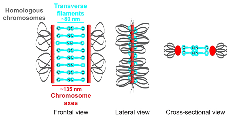
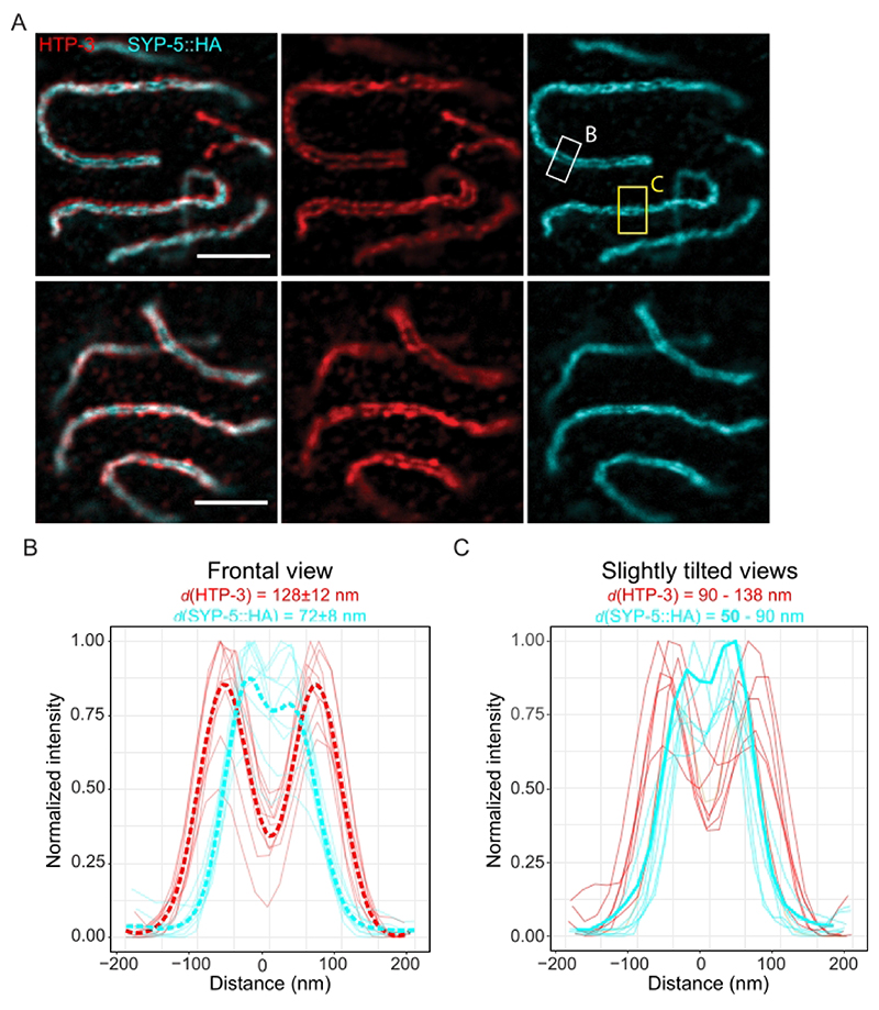
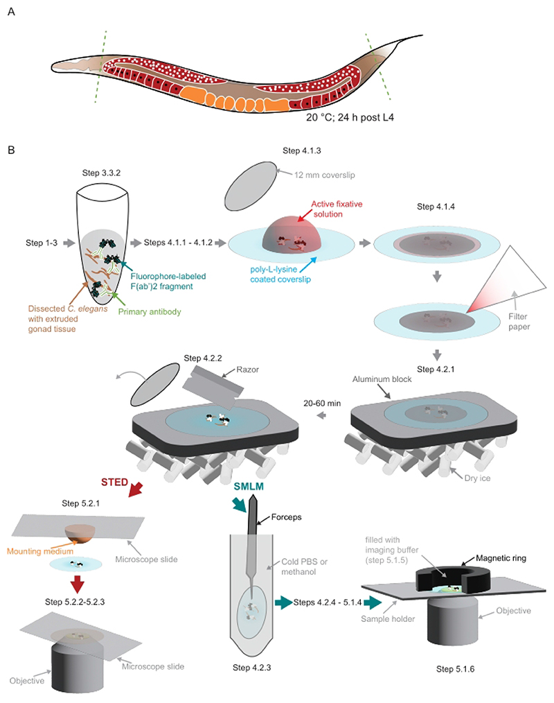
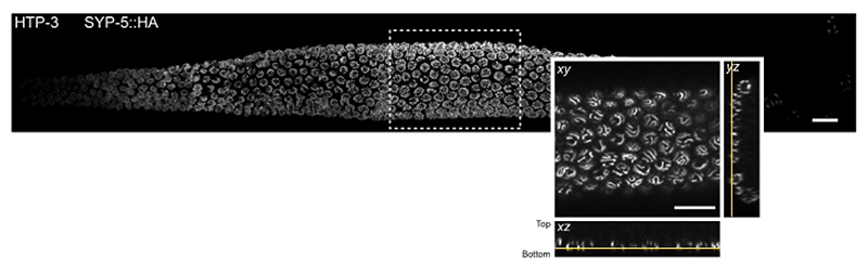
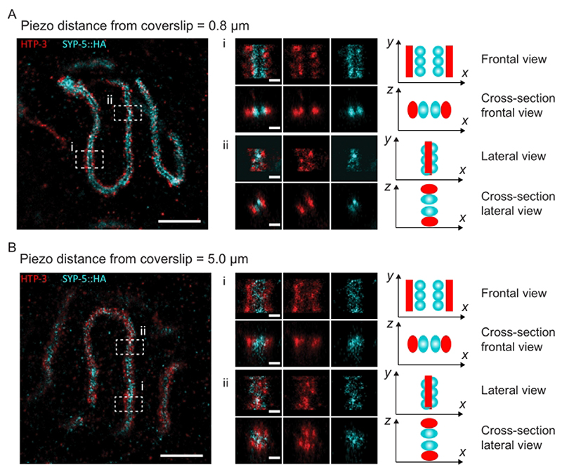
During meiosis, homologous chromosomes must recognize and adhere to one another to allow for their correct segregation. One of the key events that secures the interaction of homologous chromosomes is the assembly of the synaptonemal complex (SC) in meiotic prophase I. Even though there is little sequence homology between protein components within the SC among different species, the general structure of the SC has been highly conserved during evolution. In electron micrographs, the SC appears as a tripartite, ladder-like structure composed of lateral elements or axes, transverse filaments, and a central element. However, precisely identifying the localization of individual components within the complex by electron microscopy to determine the molecular structure of the SC remains challenging. By contrast, fluorescence microscopy allows for the identification of individual protein components within the complex. However, since the SC is only ~100 nm wide, its substructure cannot be resolved by diffraction-limited conventional fluorescence microscopy. Thus, determining the molecular architecture of the SC requires super-resolution light microscopy techniques such as structured illumination microscopy (SIM), stimulated-emission depletion (STED) microscopy, or single-molecule localization microscopy (SMLM). To maintain the structure and interactions of individual components within the SC, it is important to observe the complex in an environment that is close to its native environment in the germ cells. Therefore, we demonstrate an immunohistochemistry and imaging protocol that enables the study of the substructure of the SC in intact, extruded Caenorhabditis elegans germline tissue with SMLM and STED microscopy. Directly fixing the tissue to the coverslip reduces the movement of the samples during imaging and minimizes aberrations in the sample to achieve the high resolution necessary to visualize the substructure of the SC in its biological context.

摘要

在减数分裂过程中,同源染色体必须相互识别并结合,才能确保正确分离。确保同源染色体相互作用的一个关键事件是联会复合体 (SC) 在减数分裂前期 I 的组装。尽管不同物种的 SC 蛋白成分之间几乎没有序列同源性,但 SC 的一般结构在进化过程中高度保守。在电子显微镜下,SC 呈现出三部分、梯状结构,由侧元件或轴、横向细丝和中央元件组成。然而,通过电子显微镜精确识别复合物中各个组件的定位以确定 SC 的分子结构仍然具有挑战性。相比之下,荧光显微镜可以识别复合物中的各个蛋白成分。然而,由于 SC 只有约 100nm 宽,其亚结构无法通过传统的衍射受限荧光显微镜来解析。因此,确定 SC 的分子结构需要超分辨率光显微镜技术,如结构光照明显微镜 (SIM)、受激发射损耗 (STED) 显微镜或单分子定位显微镜 (SMLM)。为了保持 SC 中各个组件的结构和相互作用,重要的是在接近生殖细胞中其天然环境的环境中观察复合物。因此,我们展示了一种免疫组织化学和成像方案,该方案允许使用 SMLM 和 STED 显微镜研究完整的、挤出的秀丽隐杆线虫生殖系组织中 SC 的亚结构。直接将组织固定在载玻片上可以减少样品在成像过程中的运动,并最大限度地减少样品中的像差,以实现在生物背景下可视化 SC 亚结构所需的高分辨率。

https://cdn.ncbi.nlm.nih.gov/pmc/blobs/2c22/7614930/69846b9d669a/EMS155284-f006.jpg
https://cdn.ncbi.nlm.nih.gov/pmc/blobs/2c22/7614930/4a4670b2f000/EMS155284-f001.jpg
https://cdn.ncbi.nlm.nih.gov/pmc/blobs/2c22/7614930/adc830e0239e/EMS155284-f002.jpg
https://cdn.ncbi.nlm.nih.gov/pmc/blobs/2c22/7614930/7efd83acf302/EMS155284-f003.jpg
https://cdn.ncbi.nlm.nih.gov/pmc/blobs/2c22/7614930/88ae4f730eda/EMS155284-f004.jpg
https://cdn.ncbi.nlm.nih.gov/pmc/blobs/2c22/7614930/b0f9005c2292/EMS155284-f005.jpg
https://cdn.ncbi.nlm.nih.gov/pmc/blobs/2c22/7614930/69846b9d669a/EMS155284-f006.jpg

相似文献

1
Super-Resolution Microscopy of the Synaptonemal Complex within the Caenorhabditis elegans Germline.秀丽隐杆线虫生殖系中联会复合体的超分辨率显微镜观察。
J Vis Exp. 2022 Sep 13(187). doi: 10.3791/64363.
2
CRL4 regulates recombination and synaptonemal complex aggregation in the Caenorhabditis elegans germline.CRL4 调控秀丽隐杆线虫生殖细胞中的重组和联会复合体聚集。
PLoS Genet. 2019 Nov 18;15(11):e1008486. doi: 10.1371/journal.pgen.1008486. eCollection 2019 Nov.
3
Superresolution microscopy reveals the three-dimensional organization of meiotic chromosome axes in intact tissue.超分辨率显微镜技术揭示了完整组织中减数分裂染色体轴的三维结构。
Proc Natl Acad Sci U S A. 2017 Jun 13;114(24):E4734-E4743. doi: 10.1073/pnas.1702312114. Epub 2017 May 30.
4
CRA-1 uncovers a double-strand break-dependent pathway promoting the assembly of central region proteins on chromosome axes during C. elegans meiosis.CRA-1揭示了一种依赖双链断裂的途径,该途径在秀丽隐杆线虫减数分裂期间促进中央区域蛋白在染色体轴上的组装。
PLoS Genet. 2008 Jun 6;4(6):e1000088. doi: 10.1371/journal.pgen.1000088.
5
Structured illumination microscopy imaging reveals localization of replication protein A between chromosome lateral elements during mammalian meiosis.结构照明显微镜成像揭示了复制蛋白 A 在哺乳动物减数分裂过程中在染色体侧元件之间的定位。
Exp Mol Med. 2018 Aug 28;50(8):1-12. doi: 10.1038/s12276-018-0139-5.
6
A yeast two-hybrid screen for SYP-3 interactors identifies SYP-4, a component required for synaptonemal complex assembly and chiasma formation in Caenorhabditis elegans meiosis.一项针对SYP-3相互作用蛋白的酵母双杂交筛选鉴定出了SYP-4,它是秀丽隐杆线虫减数分裂中联会复合体组装和交叉形成所必需的一个组分。
PLoS Genet. 2009 Oct;5(10):e1000669. doi: 10.1371/journal.pgen.1000669. Epub 2009 Oct 2.
7
The Puf RNA-binding proteins FBF-1 and FBF-2 inhibit the expression of synaptonemal complex proteins in germline stem cells.Puf RNA 结合蛋白 FBF-1 和 FBF-2 抑制生殖干细胞中联会复合体蛋白的表达。
Development. 2010 Jun;137(11):1787-98. doi: 10.1242/dev.050799. Epub 2010 Apr 28.
8
Solving a meiotic LEGO puzzle: transverse filaments and the assembly of the synaptonemal complex in Caenorhabditis elegans.解决减数分裂的乐高难题:横向细丝和 Caenorhabditis elegans 中联会复合体的组装。
Genetics. 2011 Oct;189(2):405-9. doi: 10.1534/genetics.111.134197.
9
Organization of the synaptonemal complex during meiosis in Caenorhabditis elegans.线虫减数分裂过程中联会复合体的组织。
Genetics. 2011 Oct;189(2):411-21. doi: 10.1534/genetics.111.132431. Epub 2011 Aug 11.
10
SUMO localizes to the central element of synaptonemal complex and is required for the full synapsis of meiotic chromosomes in budding yeast.SUMO 定位于联会复合体的中央元件,并且对于芽殖酵母减数分裂染色体的完全联会是必需的。
PLoS Genet. 2013;9(10):e1003837. doi: 10.1371/journal.pgen.1003837. Epub 2013 Oct 3.

引用本文的文献

1
Skp1 proteins are structural components of the synaptonemal complex in .Skp1蛋白是……中突触复合体的结构成分。 (原句中“in”后面似乎缺失了具体信息)
Sci Adv. 2024 Feb 16;10(7):eadl4876. doi: 10.1126/sciadv.adl4876. Epub 2024 Feb 14.

本文引用的文献

1
Nanoscale structural organization and stoichiometry of the budding yeast kinetochore.出芽酵母动粒的纳米级结构组织和化学计量。
J Cell Biol. 2023 Apr 3;222(4). doi: 10.1083/jcb.202209094. Epub 2023 Jan 27.
2
Global fitting for high-accuracy multi-channel single-molecule localization.高精度多通道单分子定位的全局拟合。
Nat Commun. 2022 Jun 6;13(1):3133. doi: 10.1038/s41467-022-30719-4.
3
Architecture and Dynamics of Meiotic Chromosomes.减数分裂染色体的结构与动态。
Annu Rev Genet. 2021 Nov 23;55:497-526. doi: 10.1146/annurev-genet-071719-020235. Epub 2021 Sep 16.
4
Three-dimensional superresolution fluorescence microscopy maps the variable molecular architecture of the nuclear pore complex.三维超分辨率荧光显微镜绘制核孔复合体的可变分子结构。
Mol Biol Cell. 2021 Aug 15;32(17):1523-1533. doi: 10.1091/mbc.E20-11-0728. Epub 2021 Jun 30.
5
EMU: reconfigurable graphical user interfaces for Micro-Manager.EMU:适用于 Micro-Manager 的可重配置图形用户界面。
BMC Bioinformatics. 2020 Oct 15;21(1):456. doi: 10.1186/s12859-020-03727-8.
6
SMAP: a modular super-resolution microscopy analysis platform for SMLM data.SMAP:用于单分子定位显微镜(SMLM)数据的模块化超分辨率显微镜分析平台。
Nat Methods. 2020 Sep;17(9):870-872. doi: 10.1038/s41592-020-0938-1.
7
Optimizing imaging speed and excitation intensity for single-molecule localization microscopy.优化单分子定位显微镜的成像速度和激发强度。
Nat Methods. 2020 Sep;17(9):909-912. doi: 10.1038/s41592-020-0918-5. Epub 2020 Aug 17.
8
Tracking down the molecular architecture of the synaptonemal complex by expansion microscopy.通过扩展显微镜技术追踪联会复合体的分子结构。
Nat Commun. 2020 Jun 26;11(1):3222. doi: 10.1038/s41467-020-17017-7.
9
Multivalent weak interactions between assembly units drive synaptonemal complex formation.多价弱相互作用在组装单元之间驱动联会复合体的形成。
J Cell Biol. 2020 May 4;219(5). doi: 10.1083/jcb.201910086.
10
Identification of novel synaptonemal complex components in C. elegans.鉴定秀丽隐杆线虫中的新型联会复合体成分。
J Cell Biol. 2020 May 4;219(5). doi: 10.1083/jcb.201910043.