Antoci Valentin, Barrett Caitlin, Glasser Jillian, Barrett Thomas, Garcia Dioscaris
Department of Orthopedic Surgery, University Orthopedics Inc., East Providence, Rhode Island, United States.
Department of Orthopaedics, Warren Alpert Medical School of Brown University, Providence, Rhode Island, United States.
J Orthop Case Rep. 2022 Feb;12(2):9-13. doi: 10.13107/jocr.2022.v12.i02.2644.
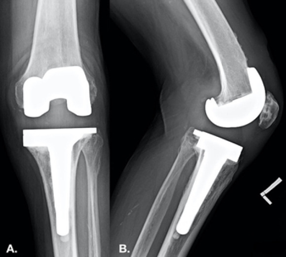
Knee pain and osteoarthritis are frequent patient complaints, with a rapidly increasing prevalence. By comparison, the prevalence of rheumatoid arthritis (RA) is significantly lower at around 1%. Inflammatory arthropathies, like RA, are difficult to differentiate from infection, crystal arthropathies, or malignancy. In addition, radiography and roentgenograms are often inconclusive or non-specific, making it much more difficult to evaluate, diagnose, and manage this condition. The current case is unique due to its location in the knee joint, rather than more common presentations in the upper extremities, and use of MRI imaging for diagnosis of RA with tenosynovitis.
In a Caucasian 70-year-old female with sudden debilitating knee pain and a large atraumatic defect over tibial plateau, MRI showed a large fluid collection within the left gracilis muscle. Gram stain and culture of the aspirate remained negative. The only significant history involved a possible diagnosis of RA.
While rheumatoid tenosynovitis is common in the upper extremities, lower extremity features have not been well reported before. We diagnosed the patient with progressive RA and rheumatoid tenosynovitis. This unique presentation and rare usage of MRI imaging may be contributing to an underreporting of this diagnosis in the lower extremities.
膝关节疼痛和骨关节炎是患者常见的主诉,其患病率正在迅速上升。相比之下,类风湿关节炎(RA)的患病率显著较低,约为1%。像RA这样的炎性关节病很难与感染、晶体性关节病或恶性肿瘤相区分。此外,X线摄影和X光片往往结论不明确或缺乏特异性,这使得评估、诊断和处理这种疾病变得更加困难。本病例因其位于膝关节,而非上肢更常见的表现,以及使用MRI成像诊断伴有腱鞘炎的RA而显得独特。
一名70岁白种女性,突发膝关节疼痛且胫骨平台出现一个无创伤的大缺损,MRI显示左侧股薄肌内有大量积液。抽吸物的革兰氏染色和培养结果均为阴性。唯一重要的病史是可能诊断为RA。
虽然类风湿性腱鞘炎在上肢很常见,但此前下肢的特征报道较少。我们诊断该患者为进展性RA和类风湿性腱鞘炎。这种独特的表现以及MRI成像的罕见使用可能导致下肢这种诊断的报告不足。