Division of Anatomy, MIC, Medical University Vienna, Vienna, Austria.
Clinical Laboratory for Bionic Extremity Reconstruction, Department of Plastic, Reconstructive and Aesthetic Surgery, Medical University of Vienna, Vienna, Austria.
Histochem Cell Biol. 2023 Jan;159(1):23-45. doi: 10.1007/s00418-022-02154-5. Epub 2022 Oct 6.
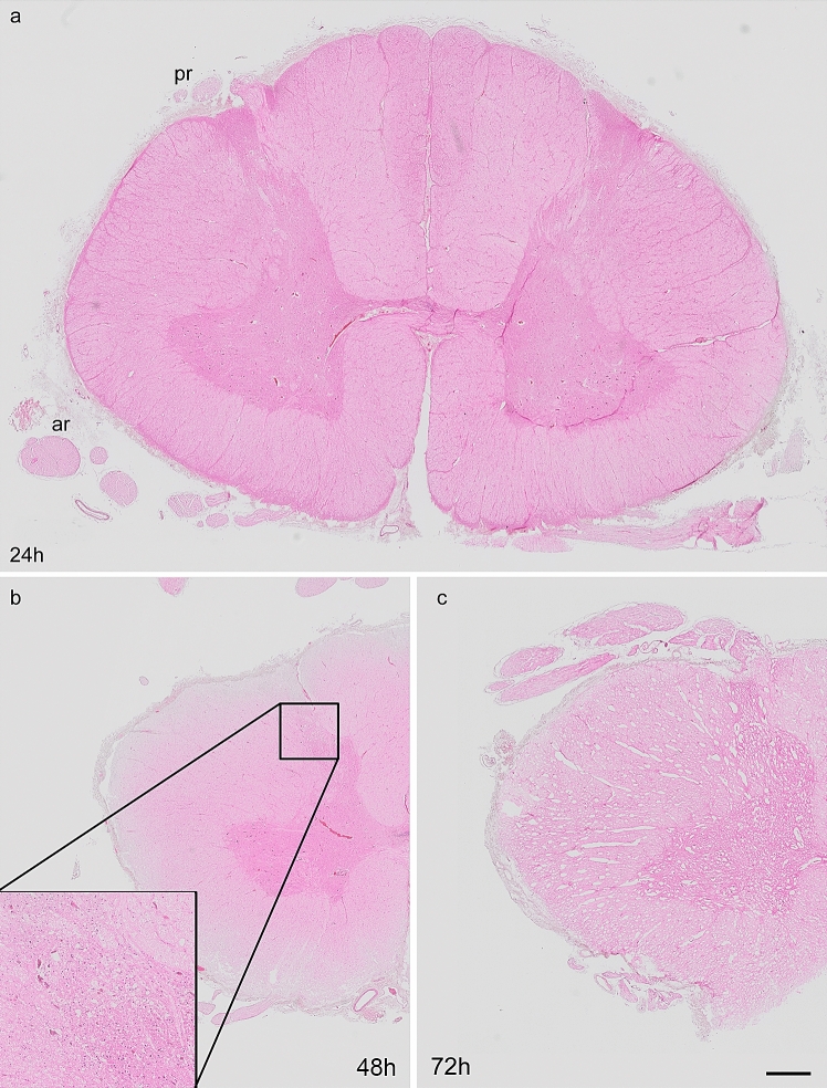
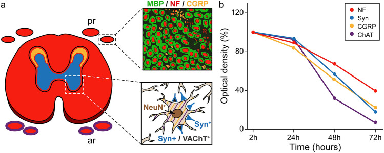
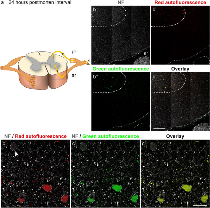
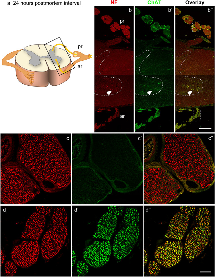
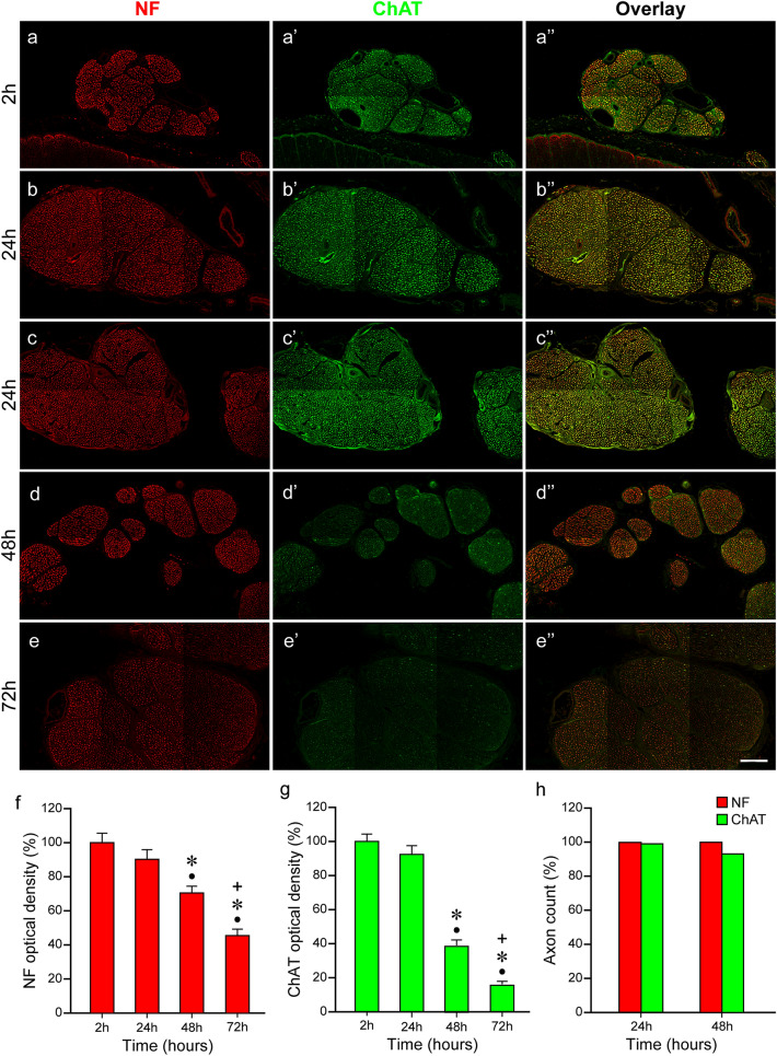
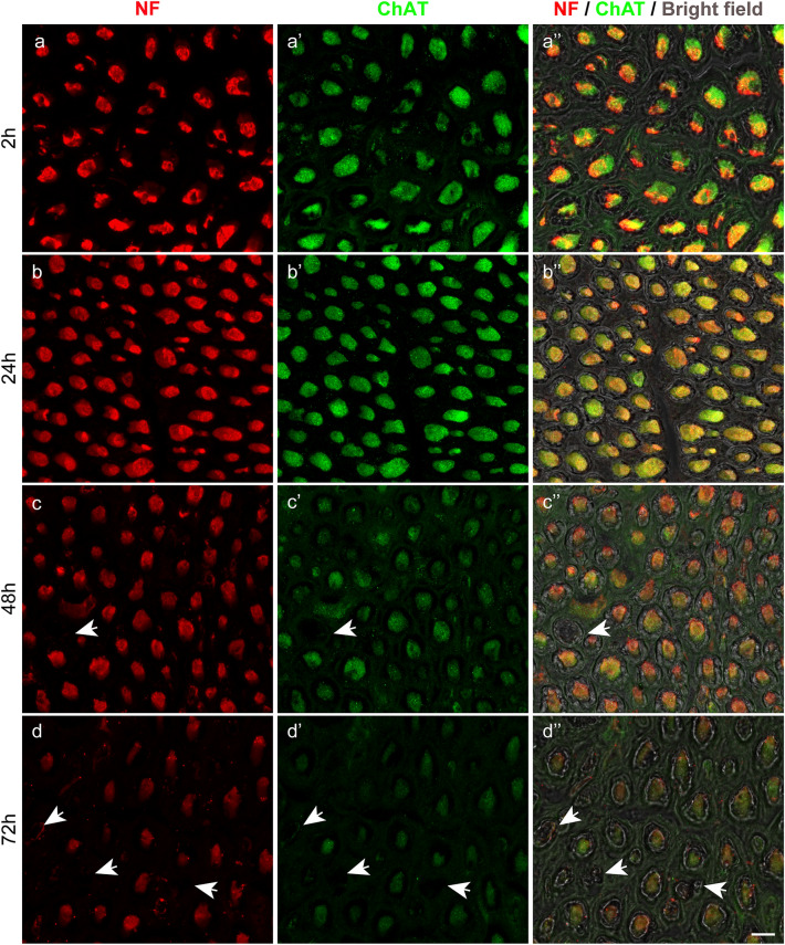
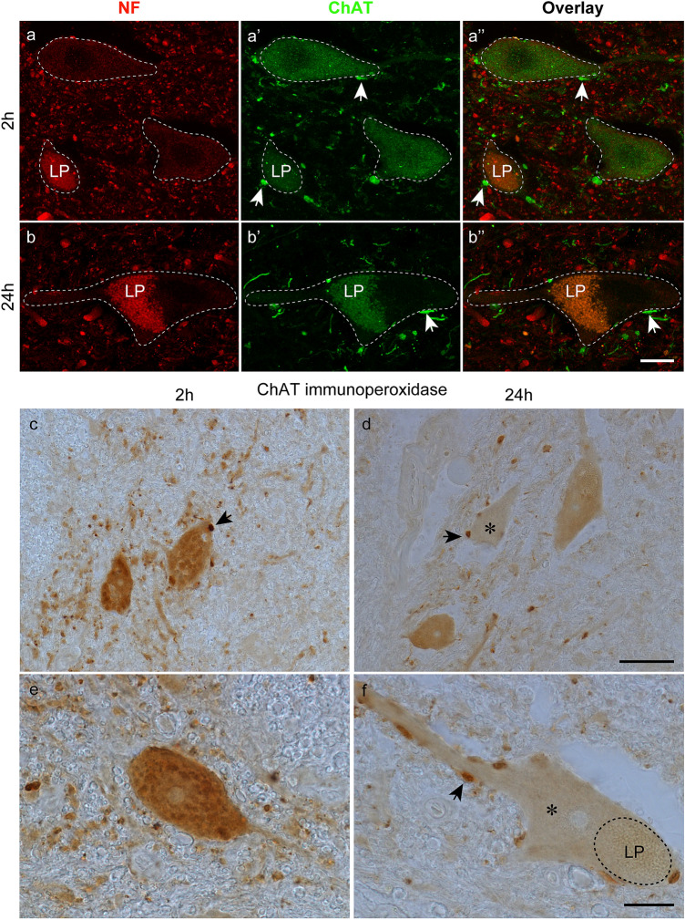
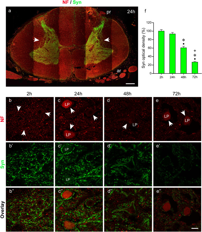
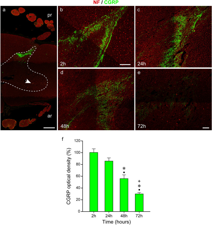
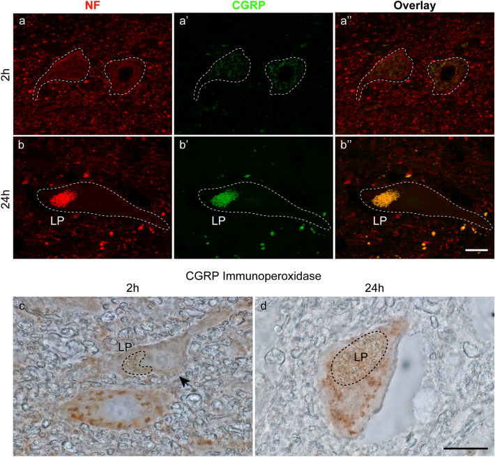
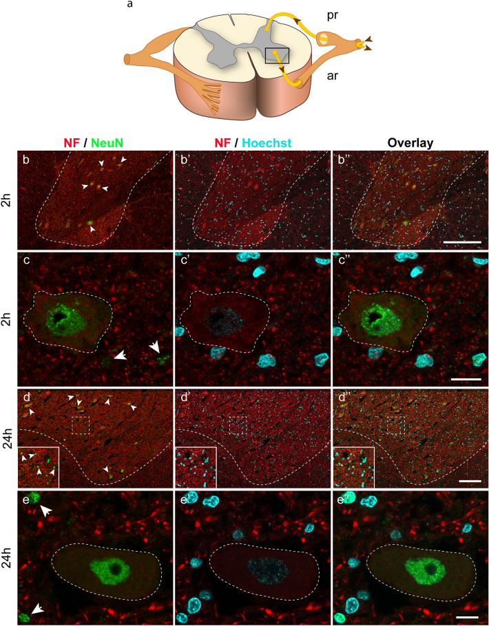
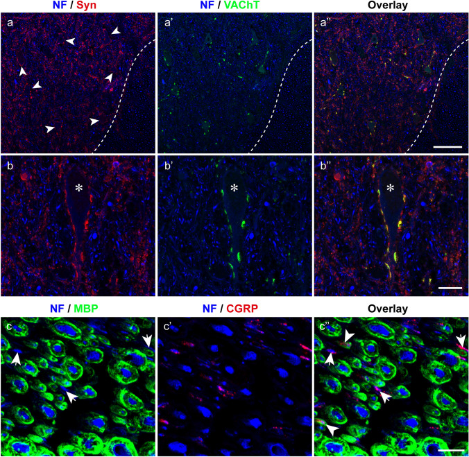
Immunohistochemistry is a powerful tool for studying neuronal tissue from humans at the molecular level. Obtaining fresh neuronal tissue from human organ donors is difficult and sometimes impossible. In anatomical body donations, neuronal tissue is dedicated to research purposes and because of its easier availability, it may be an alternative source for research. In this study, we harvested spinal cord from a single organ donor 2 h (h) postmortem and spinal cord from body donors 24, 48, and 72 h postmortem and tested how long after death, valid multi-color immunofluorescence or horseradish peroxidase (HRP) immunohistochemistry is possible. We used general and specific neuronal markers and glial markers for immunolabeling experiments. Here we showed that it is possible to visualize molecularly different neuronal elements with high precision in the body donor spinal cord 24 h postmortem and the quality of the image data was comparable to those from the fresh organ donor spinal cord. High-contrast multicolor images of the 24-h spinal cords allowed accurate automated quantification of different neuronal elements in the same sample. Although there was antibody-specific signal reduction over postmortem intervals, the signal quality for most antibodies was acceptable at 48 h but no longer at 72 h postmortem. In conclusion, our study has defined a postmortem time window of more than 24 h during which valid immunohistochemical information can be obtained from the body donor spinal cord. Due to the easier availability, neuronal tissue from body donors is an alternative source for basic and clinical research.
免疫组织化学是研究人类神经元组织的分子水平的有力工具。从人类器官捐献者中获得新鲜的神经元组织是困难的,有时甚至是不可能的。在解剖学尸体捐献中,神经元组织专门用于研究目的,由于其更容易获得,因此可能是研究的替代来源。在这项研究中,我们从单个器官捐献者死后 2 小时(h)收获脊髓,从尸体捐献者死后 24、48 和 72 小时收获脊髓,并测试死后多长时间可以进行有效的多色免疫荧光或辣根过氧化物酶(HRP)免疫组织化学。我们使用了一般和特定的神经元标记物和神经胶质标记物进行免疫标记实验。在这里,我们表明在尸体捐献者脊髓死后 24 小时可以精确地可视化分子上不同的神经元成分,并且图像数据的质量与新鲜器官捐献者脊髓的质量相当。24 小时脊髓的高对比度多色图像允许在同一样本中准确地自动定量不同的神经元成分。尽管在死后间隔期间存在抗体特异性信号降低,但大多数抗体的信号质量在 48 小时时仍然可以接受,但在 72 小时后就不再可以接受。总之,我们的研究定义了一个超过 24 小时的死后时间窗口,在此期间可以从尸体捐献者的脊髓中获得有效的免疫组织化学信息。由于更容易获得,尸体捐献者的神经元组织是基础和临床研究的替代来源。