Koehler Daniel, Sauer Markus, Karimzadeh Amir, Apostolova Ivayla, Klutmann Susanne, Adam Gerhard, Knipper Sophie, Maurer Tobias, Berliner Christoph
Department of Diagnostic and Interventional Radiology and Nuclear Medicine, University Medical Center Hamburg-Eppendorf, Martinistraße 52, 20246, Hamburg, Germany.
Martini-Klinik Prostate Cancer Center, University Hospital Hamburg-Eppendorf, Hamburg, Germany.
EJNMMI Res. 2022 Oct 9;12(1):66. doi: 10.1186/s13550-022-00938-3.
PSMA PET/CT is the recommended imaging test in cases with prostate-specific antigen (PSA) recurrence after primary therapy of prostate cancer (PCa). However, imaging protocols remain a topic of active research. The aim of the presented study was to examine the impact of additional late scans of the pelvis in [ Ga]Ga-PSMA-I&T PET/CT of patients with rising PSA after prostatectomy.
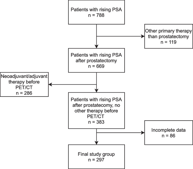
A total of 297 patients (median PSA 0.35 ng/ml, interquartile range (IQR) 0.2-0.8) who underwent early whole-body [ Ga]Ga-PSMA-I&T PET/CT (median dose 141 MBq, IQR 120-163; median 86 min, IQR 56-107) and additional late scans of the pelvis (median 180 min, IQR 170-191) were investigated retrospectively. Early and late images were staged separately according to the PROMISE criteria and compared with a final consensus of both. Standardized uptake values were analyzed for early and late scans.
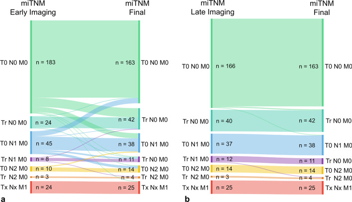
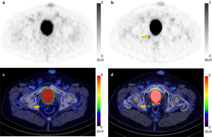
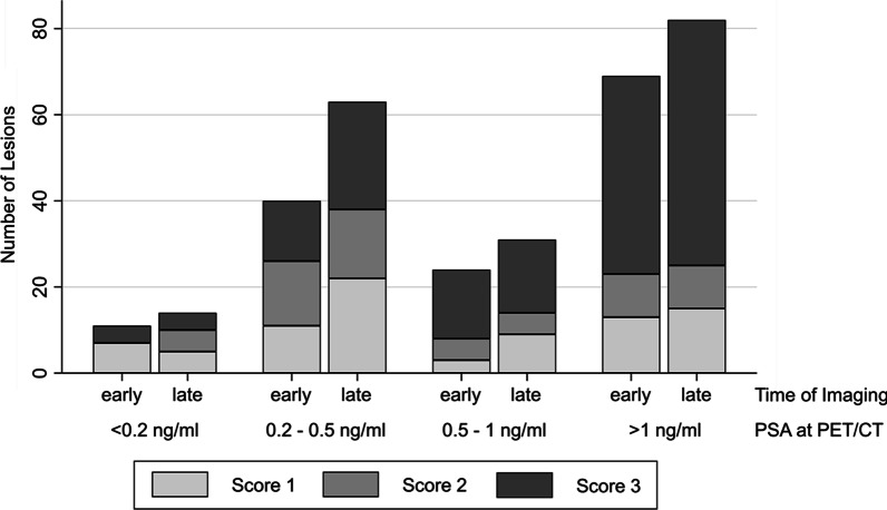
One hundred and thirty-four (45.1%) [ Ga]Ga-PSMA-I&T PET/CT showed evidence of recurrent PCa (114/38.4% early, 131/44.1% late). Of 195 lesions, 144 (73.8%) were identified correctly on early scans. 191 (97.9%) lesions were detected on late imaging. The lesion SUVmax (median 3.4, IQR 0.4-6.5 vs. median 3.9, IQR 2.6-8.2) as well as the SUVmax to background ratio (median 9.4, IQR 1.7-19.1 vs. median 15.5, IQR 9.6-34.1) increased significantly between the imaging time points (p < 0.01, respectively). Compared to the final consensus, the miTNM-staging of early scans changed in 58 (19.5%) cases. Of these, 31 patients (10.4%) with negative early scans (T0 N0 M0) were upstaged. Twenty-seven (9.1%) patients with PCa characteristic lesions on early imaging (> T0 N0 M0) were up- and/or downstaged. In 4 (1.3%) cases, PCa-related lesions were only detectable on early PET/CT leading to upstagings of late imaging.
Additional late scans of the pelvis in [ Ga]Ga-PSMA-I&T PET/CT detected more lesions and an increasing contrast compared to early imaging. This influenced the final miTNM-staging substantially.
对于前列腺癌(PCa)初次治疗后出现前列腺特异性抗原(PSA)复发的病例,PSMA PET/CT是推荐的影像学检查。然而,成像方案仍是一个活跃的研究课题。本研究的目的是探讨在前列腺切除术后PSA升高的患者中,在[镓]Ga-PSMA-I&T PET/CT检查中增加骨盆晚期扫描的影响。
对297例患者(中位PSA 0.35 ng/ml,四分位间距(IQR)0.2 - 0.8)进行回顾性研究,这些患者接受了早期全身[镓]Ga-PSMA-I&T PET/CT检查(中位剂量141 MBq,IQR 120 - 163;中位86分钟,IQR 56 - 107)以及额外的骨盆晚期扫描(中位180分钟,IQR 170 - 191)。早期和晚期图像分别根据PROMISE标准进行分期,并与两者的最终共识进行比较。分析早期和晚期扫描的标准化摄取值。
134例(45.1%)[镓]Ga-PSMA-I&T PET/CT显示有PCa复发证据(早期114例/38.4%,晚期131例/44.1%)。在195个病灶中,早期扫描正确识别出144个(73.8%)。晚期成像检测到191个(97.9%)病灶。在不同成像时间点之间,病灶SUVmax(中位值3.4,IQR 0.4 - 6.5对比中位值3.9,IQR 2.6 - 8.2)以及SUVmax与背景比值(中位值9.4,IQR 1.7 - 19.1对比中位值15.5,IQR 9.6 - 34.1)均显著增加(p均<0.01)。与最终共识相比,早期扫描的miTNM分期在58例(19.5%)病例中发生了变化。其中,31例早期扫描为阴性(T0 N0 M0)的患者(10.4%)被上调分期。27例(9.1%)早期成像有PCa特征性病灶(>T0 N0 M0)的患者被上调和/或下调分期。在4例(1.3%)病例中,PCa相关病灶仅在早期PET/CT上可检测到,导致晚期成像上调分期。
与早期成像相比,在[镓]Ga-PSMA-I&T PET/CT中增加骨盆晚期扫描可检测到更多病灶且对比度增加。这对最终的miTNM分期有重大影响。