Al-Imam Hiba, Benetti Ana R, Tomlins Pete, Gotfredsen Klaus
Department of Odontology, Section of Oral Rehabilitation, Faculty of Health and Medical Sciences, University of Copenhagen, Copenhagen, Denmark.
Department of Odontology, Section of Dental Materials, Faculty of Health and Medical Sciences, University of Copenhagen, Copenhagen, Denmark.
Biomater Investig Dent. 2022 Sep 29;9(1):84-91. doi: 10.1080/26415275.2022.2122468. eCollection 2022.
To evaluate the marginal and internal fit of lithium disilicate and zirconia crowns using two optical coherence tomography (OCT) systems in order to estimate inter-system variations.
Ten lithium disilicate and 10 cubic stabilized zirconia crowns were placed on prepared artificial teeth without cement. Marginal discrepancy and internal cement gap of the crowns were assessed on images obtained using a swept source OCT (SS-OCT) and a spectral domain OCT (SD-OCT). Medians and interquartile ranges were calculated for both materials and OCT systems. Thereafter, Wilcoxon signed rank test was carried out.
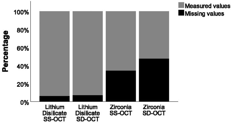
No significant difference was found between the two OCT systems for absolute marginal discrepancy of either lithium disilicate (SS-OCT: 182 µm, SD-OCT: 214 µm; = .922) or zirconia crowns (SS-OCT: 116 µm, SD-OCT: 121 µm; = .232). Regarding internal cement gap, no significant difference was found between the two OCT systems for lithium disilicate crowns (SS-OCT: 128 µm, SD-OCT: 128 µm; = .064). However, for zirconia crowns the SD-OCT showed significantly higher ( = .027) internal cement gap (92 µm) than the SS-OCT (68 µm). Moreover, it was not possible to assess the internal fit of zirconia crowns in 47% and 34% of the sites using SD-OCT and SS-OCT, respectively.
No significant difference was noted in the ability of SS-OCT and SD-OCT to assess the marginal and internal fit of lithium disilicate crowns, nor the marginal fit of zirconia crowns. On the contrary, drawbacks regarding the assessment of internal fit of zirconia crowns using both OCT systems were observed.
使用两种光学相干断层扫描(OCT)系统评估二硅酸锂和氧化锆全冠的边缘适合性和内部适合性,以估计系统间差异。
将10个二硅酸锂全冠和10个立方晶相稳定氧化锆全冠放置在未粘结水门汀的预备人工牙上。使用扫频源OCT(SS - OCT)和谱域OCT(SD - OCT)获取的图像评估全冠的边缘差异和内部水门汀间隙。计算两种材料和两种OCT系统的中位数和四分位间距。此后,进行Wilcoxon符号秩检验。
对于二硅酸锂全冠(SS - OCT:182 µm,SD - OCT:214 µm;P = 0.922)或氧化锆全冠(SS - OCT:116 µm,SD - OCT:121 µm;P = 0.232),两种OCT系统在绝对边缘差异方面未发现显著差异。关于内部水门汀间隙,对于二硅酸锂全冠,两种OCT系统之间未发现显著差异(SS - OCT:128 µm,SD - OCT:128 µm;P = 0.064)。然而,对于氧化锆全冠,SD - OCT显示的内部水门汀间隙(92 µm)显著高于SS - OCT(68 µm)(P = 0.027)。此外,分别使用SD - OCT和SS - OCT时,在47%和34%的部位无法评估氧化锆全冠的内部适合性。
在评估二硅酸锂全冠的边缘和内部适合性以及氧化锆全冠的边缘适合性方面,SS - OCT和SD - OCT的能力未发现显著差异。相反,观察到使用两种OCT系统评估氧化锆全冠内部适合性存在缺陷。