• 文献检索
  • 文档翻译
  • 深度研究
  • 学术资讯
  • Suppr Zotero 插件Zotero 插件
  • 邀请有礼
  • 套餐&价格
  • 历史记录
应用&插件
Suppr Zotero 插件Zotero 插件浏览器插件Mac 客户端Windows 客户端微信小程序
定价
高级版会员购买积分包购买API积分包
服务
文献检索文档翻译深度研究API 文档MCP 服务
关于我们
关于 Suppr公司介绍联系我们用户协议隐私条款
关注我们

Suppr 超能文献

核心技术专利:CN118964589B侵权必究
粤ICP备2023148730 号-1Suppr @ 2026

文献检索

告别复杂PubMed语法,用中文像聊天一样搜索,搜遍4000万医学文献。AI智能推荐,让科研检索更轻松。

立即免费搜索

文件翻译

保留排版,准确专业,支持PDF/Word/PPT等文件格式,支持 12+语言互译。

免费翻译文档

深度研究

AI帮你快速写综述,25分钟生成高质量综述,智能提取关键信息,辅助科研写作。

立即免费体验

基于概率性纤维束成像预测胶质母细胞瘤的真实范围。

Predicting the true extent of glioblastoma based on probabilistic tractography.

作者信息

Kis David, Szivos Laszlo, Rekecki Mark, Shukir Bayan Salam, Mate Adrienn, Hideghety Katalin, Barzo Pal

机构信息

Department of Neurosurgery, Faculty of Medicine, University of Szeged, Szeged, Hungary.

Department of Oncology, Faculty of Medicine, University of Szeged, Szeged, Hungary.

出版信息

Front Neurosci. 2022 Sep 21;16:886465. doi: 10.3389/fnins.2022.886465. eCollection 2022.

DOI:10.3389/fnins.2022.886465
PMID:36213748
原文链接:https://pmc.ncbi.nlm.nih.gov/articles/PMC9533086/
Abstract

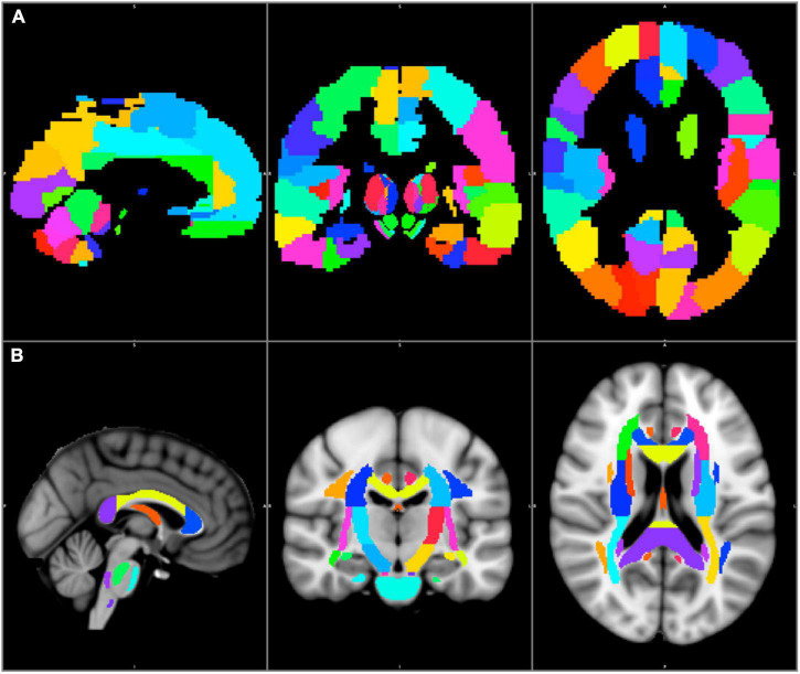
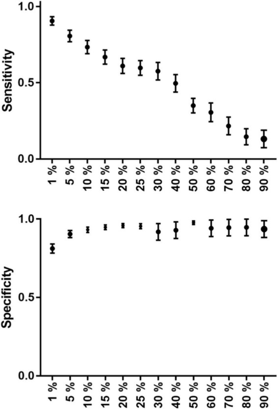
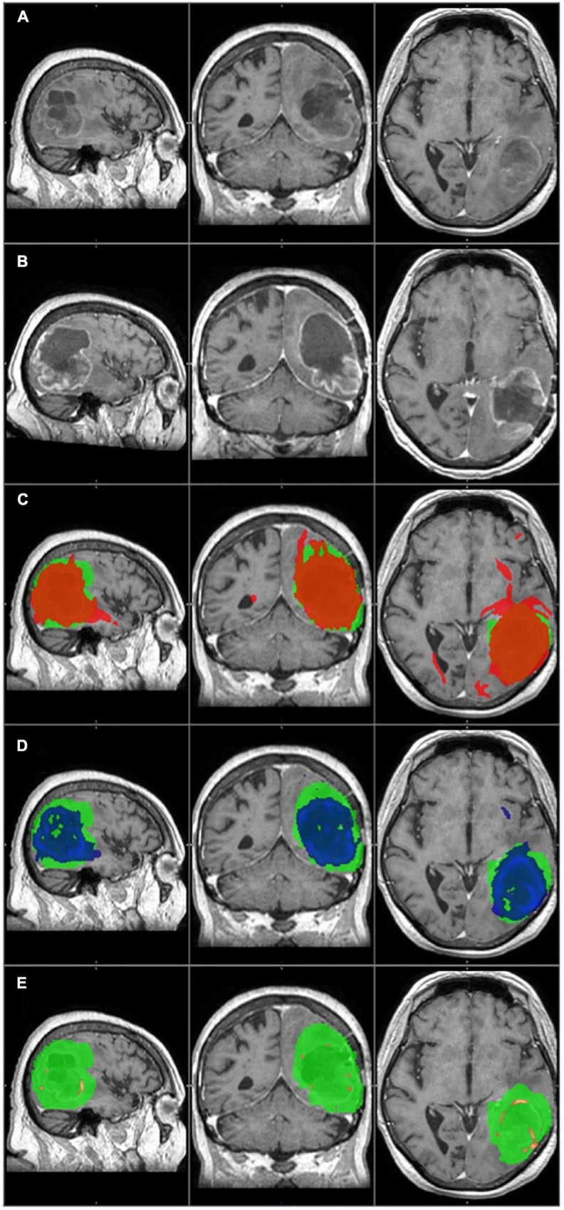
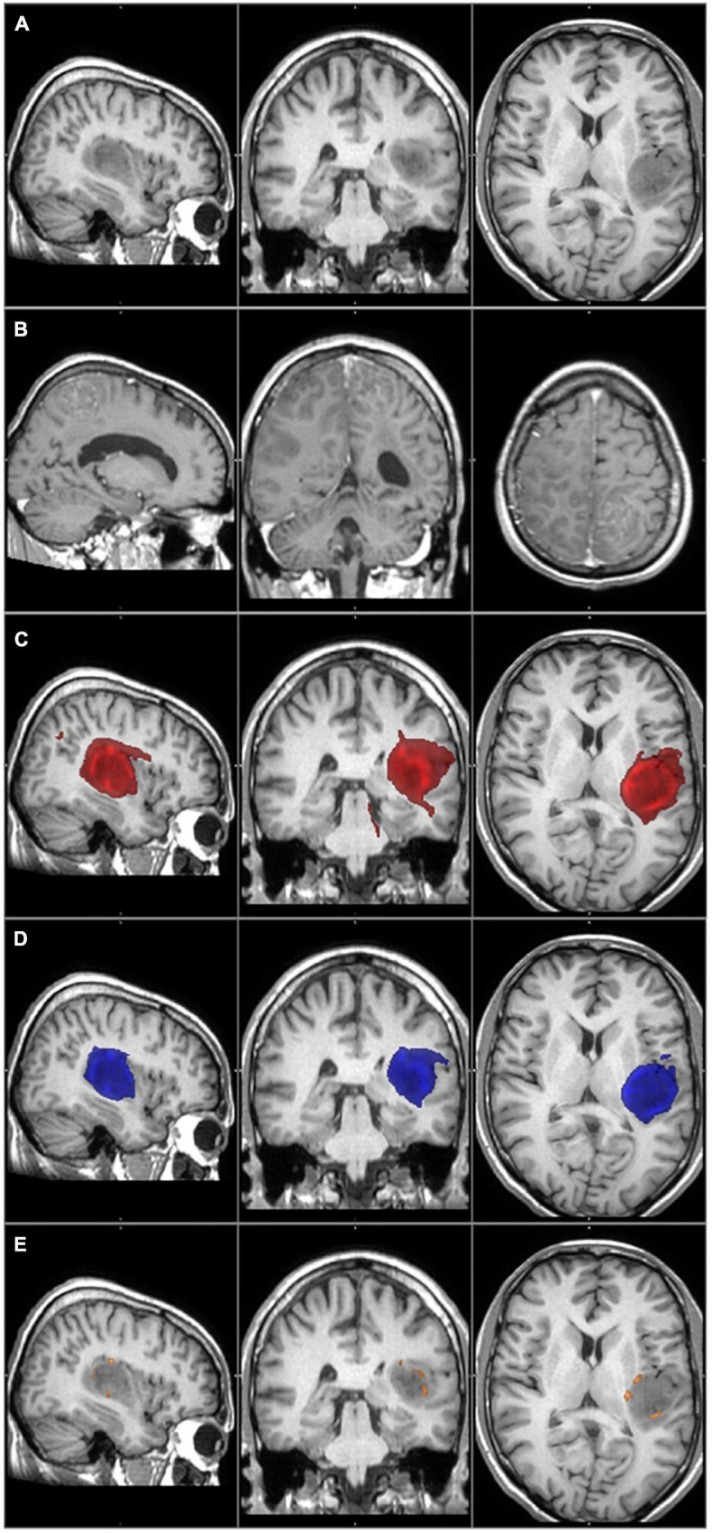
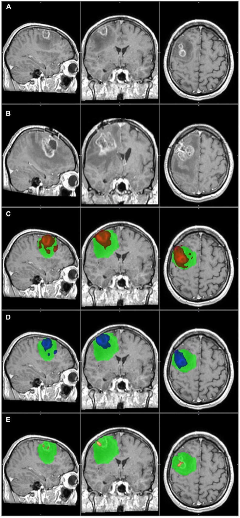
Glioblastoma is the most frequent type of primary brain tumors. Despite the advanced therapy, most of the patients die within 2 years after the diagnosis. The tumor has a typical appearance on MRI: a central hypointensity surrounded by an inhomogeneous, ring-shaped contrast enhancement along its border. Too small to be recognized by MRI, detached individual tumor cells migrate along white matter fiber tracts several centimeters away from the edge of the tumor. Usually these cells are the source of tumor recurrence. If the infiltrated brain areas could be identified, longer survival time could be achieved through supratotal resection and individually planned radiation therapy. Probabilistic tractography is an advanced imaging method that can potentially be used to identify infiltrated pathways, thus the real extent of the glioblastoma. Our study consisted of twenty high grade glioma patients. Probabilistic tractography was started from the tumor. The location of tumor recurrence on follow-up MRI was considered as the primary infiltrated white matter tracts. The results of probabilistic tractography were evaluated at thirteen different thresholds. The overlap with the tumor recurrence of each threshold level was then defined to calculate the sensitivity and specificity. In the group level, sensitivity (81%) and specificity (90%) were the most reliable at 5% threshold level. There were two outliers in the study group, both with high specificity and very low sensitivity. According to our results, probabilistic tractography can help to define the true extent of the glioblastoma at the time of diagnosis with high sensitivity and specificity. Individually planned surgery and irradiation could provide a better chance of survival in these patients.

摘要

胶质母细胞瘤是最常见的原发性脑肿瘤类型。尽管有先进的治疗方法,但大多数患者在诊断后2年内死亡。该肿瘤在磁共振成像(MRI)上有典型表现:中央低信号,周围沿边界有不均匀的环形对比增强。单个肿瘤细胞脱离肿瘤主体,体积过小以至于无法被MRI识别,它们沿着白质纤维束迁移至距离肿瘤边缘数厘米远的地方。通常这些细胞是肿瘤复发的根源。如果能够识别出肿瘤浸润的脑区,通过超全切除和个体化放疗计划,患者有望获得更长的生存时间。概率性纤维束成像技术是一种先进的成像方法,它有可能用于识别浸润路径,进而明确胶质母细胞瘤的实际范围。我们的研究纳入了20例高级别胶质瘤患者。从肿瘤部位开始进行概率性纤维束成像。将随访MRI上肿瘤复发的位置视为主要的浸润白质束。在13个不同阈值下评估概率性纤维束成像的结果。然后确定每个阈值水平与肿瘤复发的重叠情况,以计算敏感性和特异性。在组水平上,在5%阈值水平时,敏感性(81%)和特异性(90%)最为可靠。研究组中有两个异常值,特异性高但敏感性极低。根据我们的研究结果,概率性纤维束成像能够以高敏感性和特异性帮助在诊断时明确胶质母细胞瘤的实际范围。个体化的手术和放疗计划可以为这些患者提供更好的生存机会。

https://cdn.ncbi.nlm.nih.gov/pmc/blobs/0283/9533086/93bcf0f94d3d/fnins-16-886465-g005.jpg
https://cdn.ncbi.nlm.nih.gov/pmc/blobs/0283/9533086/e7a37d832a82/fnins-16-886465-g001.jpg
https://cdn.ncbi.nlm.nih.gov/pmc/blobs/0283/9533086/d4ff13e412ce/fnins-16-886465-g002.jpg
https://cdn.ncbi.nlm.nih.gov/pmc/blobs/0283/9533086/2cdf89404351/fnins-16-886465-g003.jpg
https://cdn.ncbi.nlm.nih.gov/pmc/blobs/0283/9533086/f0dfd4cea887/fnins-16-886465-g004.jpg
https://cdn.ncbi.nlm.nih.gov/pmc/blobs/0283/9533086/93bcf0f94d3d/fnins-16-886465-g005.jpg

相似文献

1
Predicting the true extent of glioblastoma based on probabilistic tractography.基于概率性纤维束成像预测胶质母细胞瘤的真实范围。
Front Neurosci. 2022 Sep 21;16:886465. doi: 10.3389/fnins.2022.886465. eCollection 2022.
2
Quantifying diffusion MRI tractography of the corticospinal tract in brain tumors with deterministic and probabilistic methods.应用确定性和概率性方法对脑肿瘤皮质脊髓束的弥散 MRI 轨迹进行量化。
Neuroimage Clin. 2013 Aug 20;3:361-8. doi: 10.1016/j.nicl.2013.08.008. eCollection 2013.
3
A systematic evaluation of intraoperative white matter tract shift in pediatric epilepsy surgery using high-field MRI and probabilistic high angular resolution diffusion imaging tractography.使用高场磁共振成像和概率性高角分辨率扩散成像纤维束示踪技术对小儿癫痫手术中白质束移位进行系统评估。
J Neurosurg Pediatr. 2017 May;19(5):592-605. doi: 10.3171/2016.11.PEDS16312. Epub 2017 Mar 17.
4
Volumetric assessment of tumor infiltration of adjacent white matter based on anatomic MRI and diffusion tensor tractography.基于解剖磁共振成像和扩散张量纤维束成像的肿瘤对相邻白质浸润的体积评估。
Acad Radiol. 2007 Apr;14(4):431-6. doi: 10.1016/j.acra.2007.01.033.
5
A registration method for improving quantitative assessment in probabilistic diffusion tractography.一种用于提高概率扩散轨迹定量评估的配准方法。
Neuroimage. 2019 Apr 1;189:288-306. doi: 10.1016/j.neuroimage.2018.12.057. Epub 2019 Jan 3.
6
Diffusion tensor tractography of the arcuate fasciculus in patients with brain tumors: Comparison between deterministic and probabilistic models.脑肿瘤患者弓状束的扩散张量纤维束成像:确定性模型与概率性模型的比较
J Biomed Sci Eng. 2013 Feb;6(2):192-200. doi: 10.4236/jbise.2013.62023.
7
Neural Fiber Integrity in High- Versus Low-Grade Glioma using Probabilistic Fiber Tracking.使用概率纤维追踪技术评估高级别与低级别胶质瘤中的神经纤维完整性
Acad Radiol. 2021 Dec;28(12):1721-1732. doi: 10.1016/j.acra.2020.09.007. Epub 2020 Oct 3.
8
Predicting the Extent of Resection in Low-Grade Glioma by Using Intratumoral Tractography to Detect Eloquent Fascicles Within the Tumor.利用肿瘤内纤维束成像检测肿瘤内明确的神经束来预测低级别胶质瘤的切除范围
Neurosurgery. 2021 Jan 13;88(2):E190-E202. doi: 10.1093/neuros/nyaa463.
9
Mapping Motor Pathways in Parkinson's Disease Patients with Subthalamic Deep Brain Stimulator: A Diffusion MRI Tractography Study.使用丘脑底核深部脑刺激器对帕金森病患者运动通路进行映射:一项扩散磁共振成像纤维束示踪研究
Neurol Ther. 2022 Jun;11(2):659-677. doi: 10.1007/s40120-022-00331-1. Epub 2022 Feb 14.
10
White matter fiber tractography: why we need to move beyond DTI.白质纤维束追踪:为什么我们需要超越 DTI。
J Neurosurg. 2013 Jun;118(6):1367-77. doi: 10.3171/2013.2.JNS121294. Epub 2013 Mar 29.

引用本文的文献

1
Preoperative FLAIR images for identifying glioblastoma boundaries.用于识别胶质母细胞瘤边界的术前液体衰减反转恢复(FLAIR)图像。
BMC Med Imaging. 2025 Jul 30;25(1):302. doi: 10.1186/s12880-025-01839-2.
2
Magnetic resonance imaging in glioblastoma radiotherapy - beyond treatment adaptation.胶质母细胞瘤放疗中的磁共振成像——超越治疗适应性。
Phys Imaging Radiat Oncol. 2025 Mar 20;34:100754. doi: 10.1016/j.phro.2025.100754. eCollection 2025 Apr.
3
Predicting glioblastoma progression using MR diffusion tensor imaging: A systematic review.

本文引用的文献

1
CBTRUS Statistical Report: Primary Brain and Other Central Nervous System Tumors Diagnosed in the United States in 2014-2018.美国 2014-2018 年诊断的原发性脑和其他中枢神经系统肿瘤 CBTRUS 统计报告。
Neuro Oncol. 2021 Oct 5;23(12 Suppl 2):iii1-iii105. doi: 10.1093/neuonc/noab200.
2
The 2021 WHO Classification of Tumors of the Central Nervous System: a summary.2021 年世卫组织中枢神经系统肿瘤分类:概述。
Neuro Oncol. 2021 Aug 2;23(8):1231-1251. doi: 10.1093/neuonc/noab106.
3
Lesion-Function Analysis from Multimodal Imaging and Normative Brain Atlases for Prediction of Cognitive Deficits in Glioma Patients.
使用磁共振扩散张量成像预测胶质母细胞瘤进展:一项系统综述。
J Neuroimaging. 2025 Jan-Feb;35(1):e13251. doi: 10.1111/jon.13251.
4
Tumor-Associated Tractography Derived from High-Angular-Resolution Q-Space MRI May Predict Patterns of Cellular Invasion in Glioblastoma.源自高角分辨率Q空间MRI的肿瘤相关纤维束成像可能预测胶质母细胞瘤的细胞侵袭模式。
Cancers (Basel). 2024 Oct 30;16(21):3669. doi: 10.3390/cancers16213669.
5
Distinguishing glioblastoma progression from treatment-related changes using DTI directionality growth analysis.使用扩散张量成像(DTI)方向性生长分析区分胶质母细胞瘤进展与治疗相关变化。
Neuroradiology. 2024 Dec;66(12):2143-2151. doi: 10.1007/s00234-024-03450-8. Epub 2024 Aug 17.
6
Advancements in Diffusion MRI Tractography for Neurosurgery.神经外科弥散磁共振成像纤维束示踪技术的进展
Invest Radiol. 2024 Jan 1;59(1):13-25. doi: 10.1097/RLI.0000000000001015. Epub 2023 Sep 14.
7
Impact of Postoperative Changes in Brain Anatomy on Target Volume Delineation for High-Grade Glioma.脑解剖结构的术后变化对高级别胶质瘤靶区勾画的影响
Cancers (Basel). 2023 May 19;15(10):2840. doi: 10.3390/cancers15102840.
基于多模态成像和标准化脑图谱的病变-功能分析对胶质瘤患者认知缺陷的预测
Cancers (Basel). 2021 May 14;13(10):2373. doi: 10.3390/cancers13102373.
4
Importance and Evidence of Extent of Resection in Glioblastoma.重要性和证据的程度切除在胶质母细胞瘤。
J Neurol Surg A Cent Eur Neurosurg. 2021 Jan;82(1):75-86. doi: 10.1055/s-0040-1701635. Epub 2020 Oct 13.
5
Automated anatomical labelling atlas 3.自动解剖学标注图谱 3.
Neuroimage. 2020 Feb 1;206:116189. doi: 10.1016/j.neuroimage.2019.116189. Epub 2019 Sep 12.
6
Supratotal Resection of Glioblastoma: Is Less More?胶质母细胞瘤的超全切除:少即是多?
Surg Technol Int. 2019 Nov 10;35:432-440.
7
Evaluating the Association Between the Extent of Resection and Survival in Gliosarcoma.评估胶质肉瘤切除范围与生存率之间的关联。
Cureus. 2019 Apr 3;11(4):e4374. doi: 10.7759/cureus.4374.
8
Current Applications of Diffusion Tensor Imaging and Tractography in Intracranial Tumor Resection.扩散张量成像和纤维束成像在颅内肿瘤切除术中的当前应用
Front Oncol. 2019 May 29;9:426. doi: 10.3389/fonc.2019.00426. eCollection 2019.
9
Non-Contrast-Enhancing Tumor: A New Frontier in Glioblastoma Research.非增强肿瘤:胶质母细胞瘤研究的新前沿。
AJNR Am J Neuroradiol. 2019 May;40(5):758-765. doi: 10.3174/ajnr.A6025. Epub 2019 Apr 4.
10
Extracellular matrix differences in glioblastoma patients with different prognoses.不同预后的胶质母细胞瘤患者的细胞外基质差异
Oncol Lett. 2019 Jan;17(1):797-806. doi: 10.3892/ol.2018.9649. Epub 2018 Oct 31.