Cozzi Francesca M, Mayrand Roxanne C, Wan Yizhou, Price Stephen J
Cambridge Brain Tumour Imaging Laboratory, Division of Neurosurgery, Department of Clinical Neurosciences, Addenbrooke's Hospital, University of Cambridge, Cambridge, UK.
J Neuroimaging. 2025 Jan-Feb;35(1):e13251. doi: 10.1111/jon.13251.
Despite multimodal treatment of glioblastoma (GBM), recurrence beyond the initial tumor volume is inevitable. Moreover, conventional MRI has shortcomings that hinder the early detection of occult white matter tract infiltration by tumor, but diffusion tensor imaging (DTI) is a sensitive probe for assessing microstructural changes, facilitating the identification of progression before standard imaging. This sensitivity makes DTI a valuable tool for predicting recurrence. A systematic review was therefore conducted to investigate how DTI, in comparison to conventional MRI, can be used for predicting GBM progression.
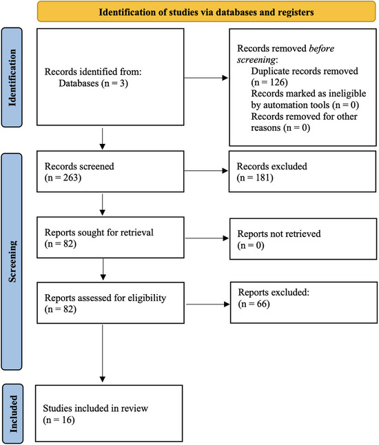
We queried three databases (PubMed, Web of Science, and Scopus) using the search terms: (diffusion tensor imaging OR DTI) AND (glioblastoma OR GBM) AND (recurrence OR progression). For included studies, data pertaining to the study type, number of GBM recurrence patients, treatment type(s), and DTI-related metrics of recurrence were extracted.
In all, 16 studies were included, from which there were 394 patients in total. Six studies reported decreased fractional anisotropy in recurrence regions, and 2 studies described the utility of connectomics/tractography for predicting tumor migratory pathways to a site of recurrence. Three studies reported evidence of tumor progression using DTI before recurrence was visible on conventional imaging.
These findings suggest that DTI metrics may be useful for guiding surgical and radiotherapy planning for GBM patients, and for informing long-term surveillance. Understanding the current state of the literature pertaining to these metrics' trends is crucial, particularly as DTI is increasingly used as a treatment-guiding imaging modality.
尽管对胶质母细胞瘤(GBM)进行了多模态治疗,但肿瘤超出初始体积复发仍不可避免。此外,传统磁共振成像(MRI)存在缺陷,阻碍了对肿瘤隐匿性白质纤维束浸润的早期检测,而扩散张量成像(DTI)是评估微观结构变化的敏感手段,有助于在标准成像显示肿瘤进展之前识别肿瘤进展情况。这种敏感性使DTI成为预测复发的有价值工具。因此,进行了一项系统评价,以研究与传统MRI相比,DTI如何用于预测GBM的进展。
我们使用搜索词“(扩散张量成像或DTI)”且“(胶质母细胞瘤或GBM)”且“(复发或进展)”查询了三个数据库(PubMed、科学网和Scopus)。对于纳入的研究,提取了与研究类型、GBM复发患者数量、治疗类型以及与DTI相关的复发指标的数据。
总共纳入了16项研究,总计394例患者。6项研究报告复发区域的各向异性分数降低,2项研究描述了连接组学/纤维束成像在预测肿瘤向复发部位迁移途径方面的作用。3项研究报告在传统成像显示复发之前,使用DTI就发现了肿瘤进展的证据。
这些发现表明,DTI指标可能有助于指导GBM患者的手术和放疗计划,并为长期监测提供信息。了解与这些指标趋势相关的文献现状至关重要,尤其是因为DTI越来越多地被用作指导治疗的成像方式。