Department of Neurochemistry, Maj Institute of Pharmacology Polish Academy of Sciences, 31-343 Krakow, Poland.
Laboratory of Pharmacogenomics, Department of Molecular Pharmacology, Maj Institute of Pharmacology Polish Academy of Sciences, 31-343 Krakow, Poland.
Int J Mol Sci. 2022 Oct 5;23(19):11817. doi: 10.3390/ijms231911817.
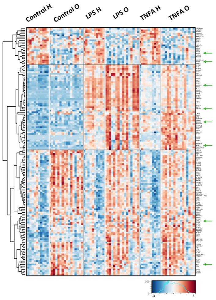
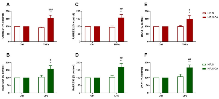
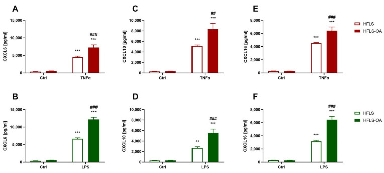
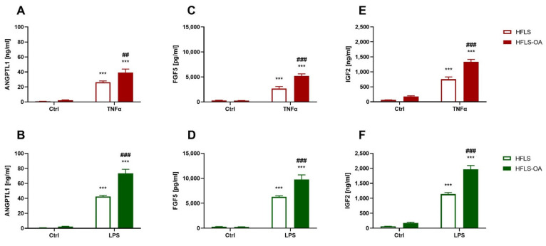
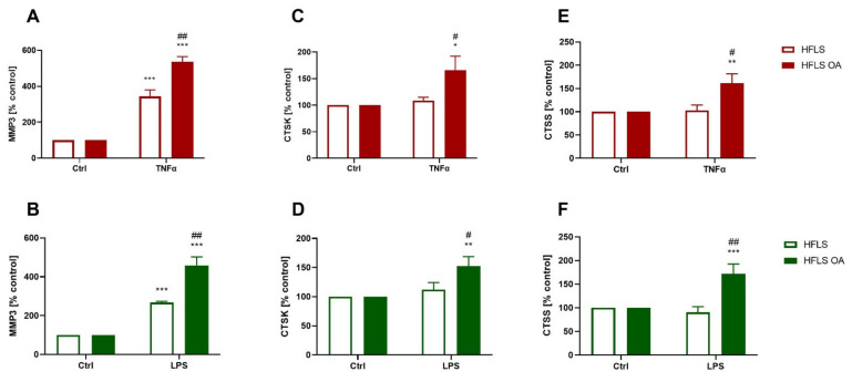
Osteoarthritis (OA) is one of the most common joint pathologies and a major cause of disability among the population of developed countries. It manifests as a gradual degeneration of the cartilage and subchondral part of the bone, leading to joint damage. Recent studies indicate that not only the cells that make up the articular cartilage but also the synoviocytes, which build the membrane surrounding the joint, contribute to the development of OA. Therefore, the aim of the study was to determine the response to inflammatory factors of osteoarthritic synoviocytes and to identify proteins secreted by them that may influence the progression of OA. This study demonstrated that fibroblast-like synoviocytes of OA patients (FLS-OA) respond more strongly to pro-inflammatory stimulation than cells obtained from control patients (FLS). These changes were observed at the transcriptome level and subsequently confirmed by protein analysis. FLS-OA stimulated by pro-inflammatory factors [such as lipopolysaccharide (LPS) and tumor necrosis factor alpha (TNFα) were shown to secrete significantly more chemokines (CXCL6, CXCL10, and CXCL16) and growth factors [angiopoietin-like protein 1 (ANGPTL1), fibroblast growth factor 5 (FGF5), and insulin-like growth factor 2 (IGF2)] than control cells. Moreover, the translation of proteolytic enzymes [matrix metalloprotease 3 (MMP3), cathepsin K (CTSK), and cathepsin S (CTSS)] by FLS-OA is increased under inflammatory conditions. Our data indicate that the FLS of OA patients are functionally altered, resulting in an enhanced response to the presence of pro-inflammatory factors in the environment, manifested by the increased production of the previously mentioned proteins, which may promote further disease progression.
骨关节炎(OA)是最常见的关节疾病之一,也是发达国家人口残疾的主要原因。它表现为软骨和骨的软骨下部分的逐渐退化,导致关节损伤。最近的研究表明,不仅构成关节软骨的细胞,而且构成关节周围膜的滑膜细胞,都有助于 OA 的发展。因此,本研究的目的是确定 OA 滑膜细胞对炎症因子的反应,并确定它们分泌的可能影响 OA 进展的蛋白质。本研究表明,OA 患者的成纤维样滑膜细胞(FLS-OA)对促炎刺激的反应比来自对照患者的细胞(FLS)更强。这些变化在转录组水平上观察到,并随后通过蛋白质分析得到证实。促炎因子(如脂多糖(LPS)和肿瘤坏死因子α(TNFα)刺激的 FLS-OA 分泌明显更多的趋化因子(CXCL6、CXCL10 和 CXCL16)和生长因子[血管生成素样蛋白 1(ANGPTL1)、成纤维细胞生长因子 5(FGF5)和胰岛素样生长因子 2(IGF2)]比对照细胞。此外,在炎症条件下,FLS-OA 翻译的蛋白水解酶[基质金属蛋白酶 3(MMP3)、组织蛋白酶 K(CTSK)和组织蛋白酶 S(CTSS)]增加。我们的数据表明,OA 患者的 FLS 功能发生改变,导致对环境中促炎因子的存在反应增强,表现为先前提到的蛋白质产生增加,这可能促进进一步的疾病进展。