Lnu Priyal, Sehgal Vineet, Bhalla Sehgal Lucky, Gulati Nihal, Kapila Saniya
Neurology, Sehgal's Neuro & Child Care Centre, Amritsar, IND.
Neurology, Lady Hardinge Medical College, Delhi, IND.
Cureus. 2022 Sep 11;14(9):e29048. doi: 10.7759/cureus.29048. eCollection 2022 Sep.
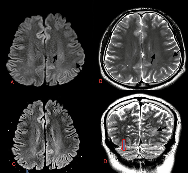
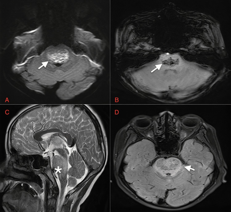
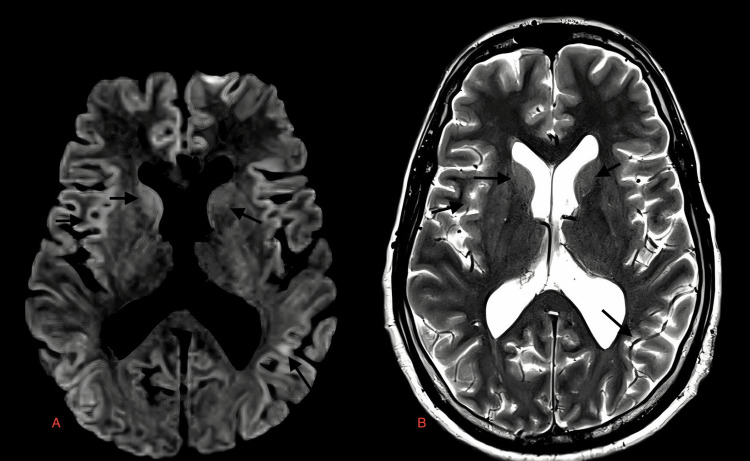
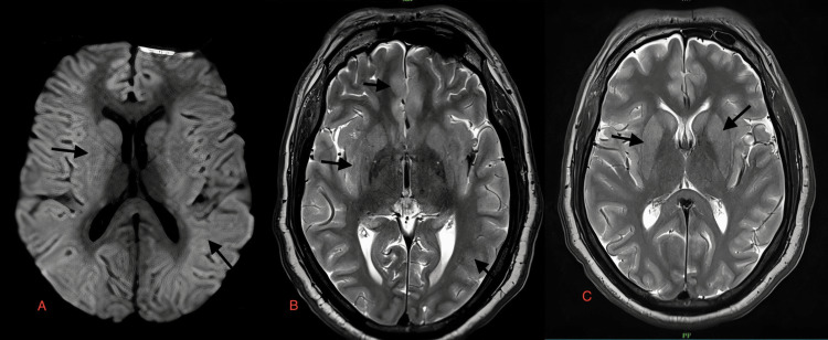
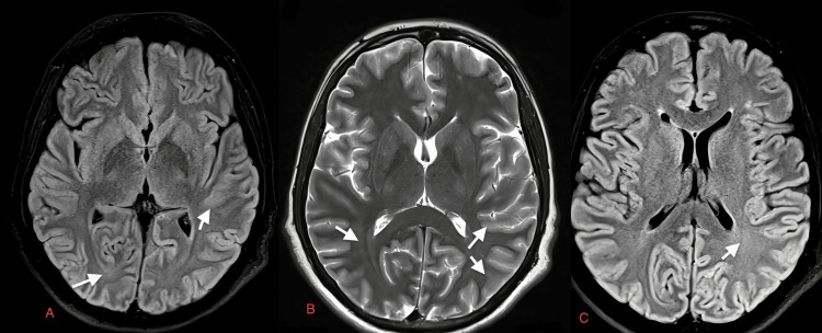
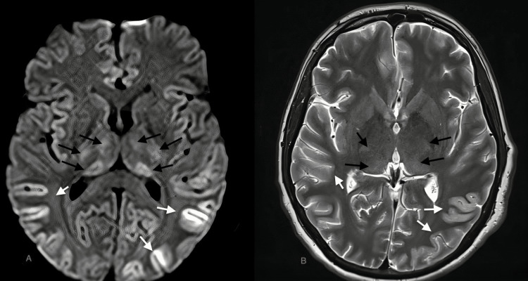
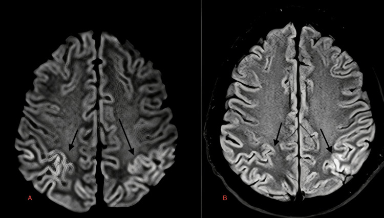
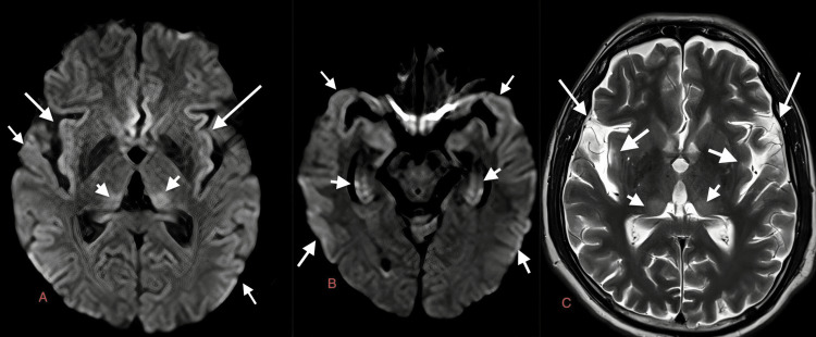
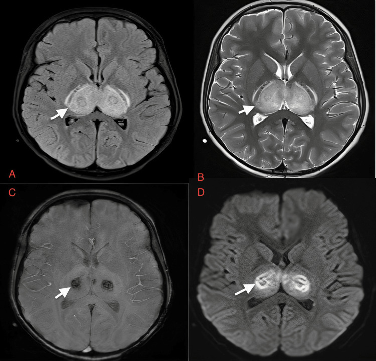
Background In this study, we aimed to describe eight cases of dengue encephalitis along with their magnetic resonance imaging (MRI) findings. Dengue encephalitis is caused by an arbovirus that has four strains DENV1-DENV4. The dengue virus is usually non-neurotropic but DENV2 & DENV3 are neurotropic. Dengue encephalitis is characterized by headaches, seizures, and altered consciousness. Methodology At our facility, we performed 3T MRI on eight suspected cases of dengue encephalitis using the criteria established by Varatharaj et al. We were able to diagnose dengue encephalitis based on the proposed criteria which included symptoms, serology, cerebrospinal fluid (CSF) analysis results, MRI findings, and routine blood laboratory workup in dengue encephalitis. Because numerous brain regions are potentially impacted in severe cases of dengue encephalitis, an MRI of the brain can reveal the severity of the condition. In deteriorating situations, it may detect whether or not further regions are being impacted. Hence, MRI should be done in all suspected cases of dengue encephalitis. Results The changes observed on MRI of the eight cases were in the supra-tentorium (deep periventricular white matter, subcortical white matter, and deep gray matter of the brain, which includes basal ganglia and thalami), infra-tentorium (cerebellar white matter and brainstem, which includes pons), and occasionally in cortical gray matter. The MRI showed mild-to-moderate hyperintensities on T2-weighted images and fluid-attenuated inversion recovery sequence (FLAIR); diffusion restriction is seen on diffusion-weighted images. The neurological clinical features included non-localizing signs and symptoms such as altered mental status, headache with vomiting, and fever. Conclusions The commonly affected areas of the brain in dengue encephalitis are the basal ganglia, thalamus, brainstem, cerebellum, cortical white matter, periventricular white matter, and cortical gray matter, which are all hyperintense on T2-weighted images and FLAIR. The lesions are iso or hypointense on T1-weighted images and micro-hemorrhages appear as blooming on susceptibility-weighted MRI. MRI is a crucial initial investigation in suspected cases of dengue encephalitis and known cases of dengue fever experiencing worsening neurological conditions.
背景 在本研究中,我们旨在描述8例登革热脑炎病例及其磁共振成像(MRI)表现。登革热脑炎由一种虫媒病毒引起,该病毒有4个毒株,即DENV1 - DENV4。登革热病毒通常不嗜神经,但DENV2和DENV3嗜神经。登革热脑炎的特征为头痛、癫痫发作和意识改变。
方法 在我们的机构,我们按照Varatharaj等人制定的标准,对8例疑似登革热脑炎病例进行了3T MRI检查。我们能够根据所提出的标准诊断登革热脑炎,这些标准包括症状、血清学、脑脊液(CSF)分析结果、MRI表现以及登革热脑炎的常规血液实验室检查。由于在严重的登革热脑炎病例中许多脑区可能受到影响,脑部MRI可以揭示病情的严重程度。在病情恶化的情况下,它可以检测是否有更多脑区受到影响。因此,对于所有疑似登革热脑炎病例都应进行MRI检查。
结果 在8例病例的MRI上观察到的变化位于幕上(脑室周围深部白质、皮质下白质以及脑深部灰质,包括基底神经节和丘脑)、幕下(小脑白质和脑干,包括脑桥),偶尔也见于皮质灰质。MRI在T2加权像和液体衰减反转恢复序列(FLAIR)上显示轻度至中度高信号;在扩散加权像上可见扩散受限。神经学临床特征包括非定位性体征和症状,如精神状态改变、头痛伴呕吐和发热。
结论 登革热脑炎中脑部常见的受累区域是基底神经节、丘脑、脑干、小脑、皮质白质、脑室周围白质和皮质灰质,这些区域在T2加权像和FLAIR上均为高信号。病变在T1加权像上呈等信号或低信号,在磁敏感加权MRI上微出血表现为磁敏感伪影。MRI是疑似登革热脑炎病例以及已知登革热发热病例出现神经状况恶化时至关重要的初步检查。