• 文献检索
  • 文档翻译
  • 深度研究
  • 学术资讯
  • Suppr Zotero 插件Zotero 插件
  • 邀请有礼
  • 套餐&价格
  • 历史记录
应用&插件
Suppr Zotero 插件Zotero 插件浏览器插件Mac 客户端Windows 客户端微信小程序
定价
高级版会员购买积分包购买API积分包
服务
文献检索文档翻译深度研究API 文档MCP 服务
关于我们
关于 Suppr公司介绍联系我们用户协议隐私条款
关注我们

Suppr 超能文献

核心技术专利:CN118964589B侵权必究
粤ICP备2023148730 号-1Suppr @ 2026

文献检索

告别复杂PubMed语法,用中文像聊天一样搜索,搜遍4000万医学文献。AI智能推荐,让科研检索更轻松。

立即免费搜索

文件翻译

保留排版,准确专业,支持PDF/Word/PPT等文件格式,支持 12+语言互译。

免费翻译文档

深度研究

AI帮你快速写综述,25分钟生成高质量综述,智能提取关键信息,辅助科研写作。

立即免费体验

视神经头部髓鞘相关蛋白、GFAP 和 Iba1 在早期至中度实验性青光眼的非人类灵长类动物中的改变。

Optic Nerve Head Myelin-Related Protein, GFAP, and Iba1 Alterations in Non-Human Primates With Early to Moderate Experimental Glaucoma.

机构信息

Optic Nerve Head Research Laboratory, Devers Eye Institute, Legacy Research Institute, Portland, Oregon, United States.

Discoveries in Sight, Devers Eye Institute, Legacy Research Institute, Portland, Oregon, United States.

出版信息

Invest Ophthalmol Vis Sci. 2022 Oct 3;63(11):9. doi: 10.1167/iovs.63.11.9.

DOI:10.1167/iovs.63.11.9
PMID:36239974
原文链接:https://pmc.ncbi.nlm.nih.gov/articles/PMC9586137/
Abstract

PURPOSE

The purpose of this study was to test if optic nerve head (ONH) myelin basic protein (MBP), 2',3'-cyclic nucleotide 3'-phosphodiesterase (CNPase), glial fibrillary acidic protein (GFAP), and ionized calcium binding adaptor molecule 1 (Iba1) proteins are altered in non-human primate (NHP) early/moderate experimental glaucoma (EG).

METHODS

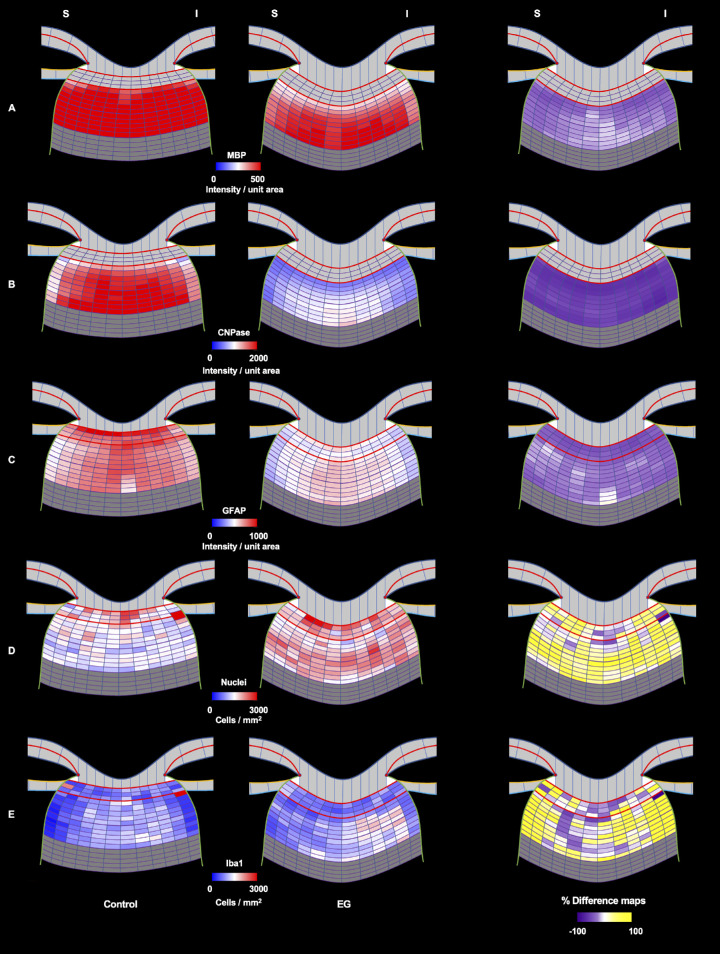
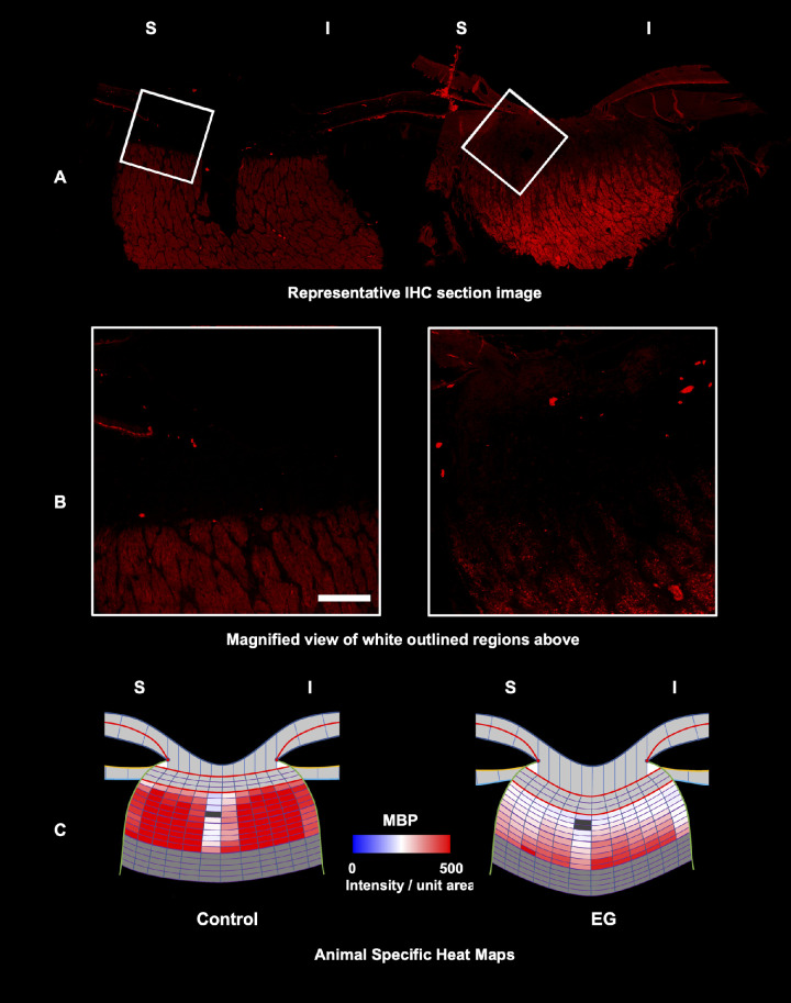
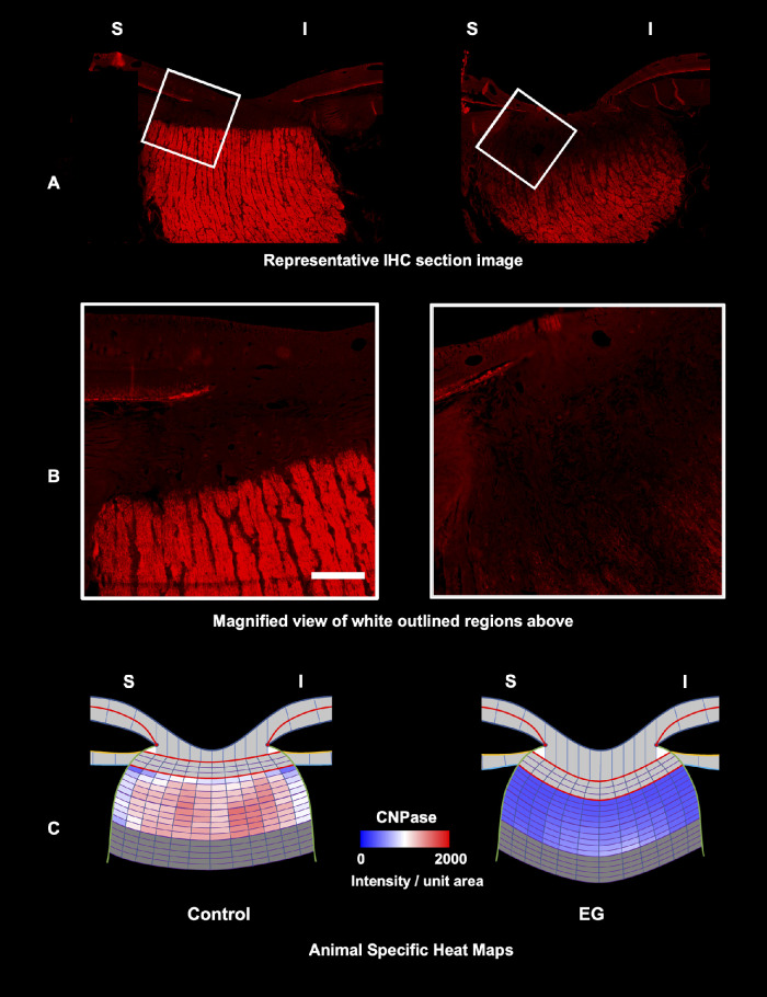
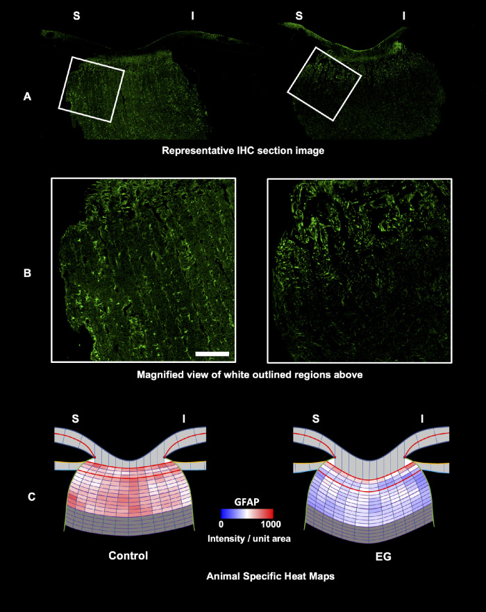
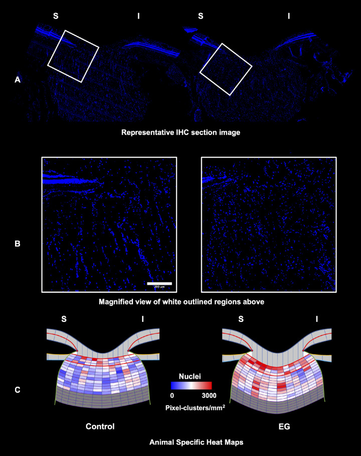
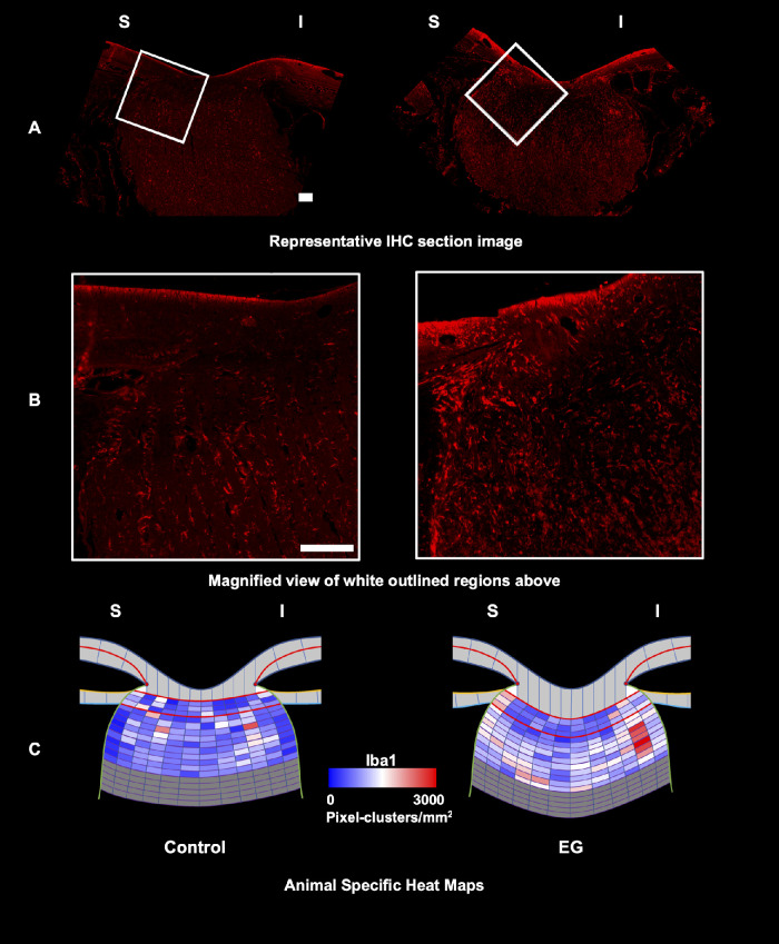
Following paraformaldehyde perfusion, control and EG eye ONH tissues from four NHPs were paraffin embedded and serially (5 µm) vertically sectioned. Anti-MBP, CNPase, GFAP, Iba1, and nuclear dye-stained sections were imaged using sub-saturating light intensities. Whole-section images were segmented creating anatomically consistent laminar (L) and retrolaminar (RL) regions/sub-regions. EG versus control eye intensity/pixel-cluster density data within L and two RL regions (RL1 [1-250 µm]/RL2 [251-500 µm] from L) were compared using random effects models within the statistical program "R."

RESULTS

EG eye retinal nerve fiber loss ranged from 0% to 20%. EG eyes' MBP and CNPase intensity were decreased within the RL1 (MBP = 31.4%, P < 0.001; CNPase =62.3%, P < 0.001) and RL2 (MBP = 19.6%, P < 0.001; CNPase = 56.1%, P = 0.0004) regions. EG eye GFAP intensity was decreased in the L (41.6%, P < 0.001) and RL regions (26.7% for RL1, and 28.4% for RL2, both P < 0.001). Iba1+ and NucBlue pixel-cluster density were increased in the laminar (28.2%, P = 0.03 and 16.6%, P = 0.008) and both RL regions (RL1 = 37.3%, P = 0.01 and 23.7%, P = 0.0002; RL2 = 53.7%, P = 0.002 and 33.2%, P < 0.001).

CONCLUSIONS

Retrolaminar myelin disruption occurs early in NHP EG and may be accompanied by laminar and retrolaminar decreases in astrocyte process labeling and increases in microglial/ macrophage density. The mechanistic and therapeutic implications of these findings warrant further study.

摘要

目的

本研究旨在检测视神经头部(ONH)髓鞘碱性蛋白(MBP)、2',3'-环核苷酸 3'-磷酸二酯酶(CNPase)、胶质纤维酸性蛋白(GFAP)和离子钙结合接头分子 1(Iba1)在非人类灵长类动物(NHP)早期/中度实验性青光眼(EG)中的变化。

方法

在进行多聚甲醛灌注后,来自四只 NHP 的对照眼和 EG 眼的 ONH 组织被石蜡包埋并进行连续(5μm)垂直切片。使用亚饱和光强度对抗 MBP、CNPase、GFAP、Iba1 和核染料染色的切片进行成像。通过在统计程序“R”中使用随机效应模型,对整个切片图像进行分割,创建解剖学上一致的层(L)和后层(RL)区域/亚区。使用随机效应模型在统计程序“R”中比较 L 和两个 RL 区域(RL1 [1-250μm]/RL2 [251-500μm])中 EG 眼与对照眼的强度/像素簇密度数据。

结果

EG 眼的视网膜神经纤维损失范围为 0%至 20%。EG 眼中 RL1(MBP=31.4%,P<0.001;CNPase=62.3%,P<0.001)和 RL2(MBP=19.6%,P<0.001;CNPase=56.1%,P=0.0004)区域的 MBP 和 CNPase 强度降低。EG 眼 GFAP 强度在 L(41.6%,P<0.001)和 RL 区域(RL1 为 26.7%,RL2 为 28.4%,均 P<0.001)降低。Iba1+和 NucBlue 像素簇密度在层状区(28.2%,P=0.03 和 16.6%,P=0.008)和两个 RL 区(RL1=37.3%,P=0.01 和 23.7%,P=0.0002;RL2=53.7%,P=0.002 和 33.2%,P<0.001)增加。

结论

在 NHP EG 中,后层髓鞘破坏发生较早,可能伴有层状和后层星形胶质细胞突起标记物减少以及小胶质细胞/巨噬细胞密度增加。这些发现的机制和治疗意义值得进一步研究。

https://cdn.ncbi.nlm.nih.gov/pmc/blobs/77d4/9586137/ff751db561f6/iovs-63-11-9-f011.jpg
https://cdn.ncbi.nlm.nih.gov/pmc/blobs/77d4/9586137/36a62d3d57e1/iovs-63-11-9-f001.jpg
https://cdn.ncbi.nlm.nih.gov/pmc/blobs/77d4/9586137/f4415a7b98b9/iovs-63-11-9-f002.jpg
https://cdn.ncbi.nlm.nih.gov/pmc/blobs/77d4/9586137/9341b0de3c47/iovs-63-11-9-f003.jpg
https://cdn.ncbi.nlm.nih.gov/pmc/blobs/77d4/9586137/df2111022f14/iovs-63-11-9-f004.jpg
https://cdn.ncbi.nlm.nih.gov/pmc/blobs/77d4/9586137/a1bcc4864c7f/iovs-63-11-9-f005.jpg
https://cdn.ncbi.nlm.nih.gov/pmc/blobs/77d4/9586137/1e5a7f143cea/iovs-63-11-9-f006.jpg
https://cdn.ncbi.nlm.nih.gov/pmc/blobs/77d4/9586137/ab5d6256e0f0/iovs-63-11-9-f007.jpg
https://cdn.ncbi.nlm.nih.gov/pmc/blobs/77d4/9586137/047398e9262b/iovs-63-11-9-f008.jpg
https://cdn.ncbi.nlm.nih.gov/pmc/blobs/77d4/9586137/f531da2d5e00/iovs-63-11-9-f009.jpg
https://cdn.ncbi.nlm.nih.gov/pmc/blobs/77d4/9586137/b6297df0ee0d/iovs-63-11-9-f010.jpg
https://cdn.ncbi.nlm.nih.gov/pmc/blobs/77d4/9586137/ff751db561f6/iovs-63-11-9-f011.jpg

相似文献

1
Optic Nerve Head Myelin-Related Protein, GFAP, and Iba1 Alterations in Non-Human Primates With Early to Moderate Experimental Glaucoma.视神经头部髓鞘相关蛋白、GFAP 和 Iba1 在早期至中度实验性青光眼的非人类灵长类动物中的改变。
Invest Ophthalmol Vis Sci. 2022 Oct 3;63(11):9. doi: 10.1167/iovs.63.11.9.
2
Hyaluronic acid in the normal and glaucomatous optic nerve.正常及青光眼视神经中的透明质酸
Exp Eye Res. 1997 Apr;64(4):587-95. doi: 10.1006/exer.1996.0245.
3
Retrolaminar Demyelination of Structurally Intact Axons in Nonhuman Primate Experimental Glaucoma.结构完整轴突的神经髓鞘在非人类灵长类实验性青光眼的后退性脱髓鞘。
Invest Ophthalmol Vis Sci. 2024 Feb 1;65(2):36. doi: 10.1167/iovs.65.2.36.
4
Neurodegeneration, Myelin Loss and Glial Response in the Three-Vessel Global Ischemia Model in Rat.大鼠三血管全脑缺血模型中的神经退行性变、髓鞘丢失和神经胶质反应。
Int J Mol Sci. 2020 Aug 28;21(17):6246. doi: 10.3390/ijms21176246.
5
Comparative quantitative study of astrocytes and capillary distribution in optic nerve laminar regions.视神经层区星形胶质细胞与毛细血管分布的比较定量研究。
Exp Eye Res. 2014 Apr;121:11-22. doi: 10.1016/j.exer.2014.02.008. Epub 2014 Feb 19.
6
Disorders in myelination in the twitcher mutant: immunohistochemical and biochemical studies.震颤突变体髓鞘形成障碍:免疫组织化学和生物化学研究
Neurochem Res. 1985 Aug;10(8):1129-41. doi: 10.1007/BF00965887.
7
3-D histomorphometry of the normal and early glaucomatous monkey optic nerve head: prelaminar neural tissues and cupping.正常和早期青光眼猴视神经乳头的三维组织形态计量学:板层前神经组织与视杯
Invest Ophthalmol Vis Sci. 2007 Nov;48(11):5068-84. doi: 10.1167/iovs.07-0790.
8
Oxidative injury to blood vessels and glia of the pre-laminar optic nerve head in human glaucoma.人类青光眼患者视盘前层视神经头血管和神经胶质的氧化损伤。
Exp Eye Res. 2008 Nov;87(5):409-14. doi: 10.1016/j.exer.2008.07.011. Epub 2008 Aug 5.
9
Expression of glial cells molecules in the optic nerve of adult dromedary camel (Camelus dromedarius): A histological and immunohistochemical analysis.成年单峰驼(骆驼属单峰驼)视神经中神经胶质细胞分子的表达:组织学和免疫组织化学分析
Anat Histol Embryol. 2019 Jan;48(1):74-86. doi: 10.1111/ahe.12413. Epub 2018 Nov 18.
10
Optic nerve head and intraocular pressure in the guinea pig eye.豚鼠眼的视神经乳头与眼压
Exp Eye Res. 2016 May;146:7-16. doi: 10.1016/j.exer.2015.12.007. Epub 2015 Dec 15.

引用本文的文献

1
Rebalancing translaminar pressure difference: a novel glaucoma surgery-a pilot trial in non-human primates.重新平衡跨板层压力差:一种新型青光眼手术——非人灵长类动物的初步试验
Int J Ophthalmol. 2025 Sep 18;18(9):1619-1625. doi: 10.18240/ijo.2025.09.02. eCollection 2025.
2
Astrocytes in the mouse brain respond bilaterally to unilateral retinal neurodegeneration.小鼠大脑中的星形胶质细胞对单侧视网膜神经变性产生双侧反应。
Proc Natl Acad Sci U S A. 2025 Mar 18;122(11):e2418249122. doi: 10.1073/pnas.2418249122. Epub 2025 Mar 10.
3
Retrolaminar Demyelination of Structurally Intact Axons in Nonhuman Primate Experimental Glaucoma.

本文引用的文献

1
Reactive astrocyte nomenclature, definitions, and future directions.反应性星形胶质细胞命名、定义和未来方向。
Nat Neurosci. 2021 Mar;24(3):312-325. doi: 10.1038/s41593-020-00783-4. Epub 2021 Feb 15.
2
When Is a Control Not a Control? Reactive Microglia Occur Throughout the Control Contralateral Pathway of Retinal Ganglion Cell Projections in Experimental Glaucoma.当控制不再是控制时?在实验性青光眼的视网膜神经节细胞投射的对照侧通路上,反应性小胶质细胞普遍存在。
Transl Vis Sci Technol. 2021 Jan 12;10(1):22. doi: 10.1167/tvst.10.1.22. eCollection 2021 Jan.
3
Thyroid hormone and thyromimetics inhibit myelin and axonal degeneration and oligodendrocyte loss in EAE.
结构完整轴突的神经髓鞘在非人类灵长类实验性青光眼的后退性脱髓鞘。
Invest Ophthalmol Vis Sci. 2024 Feb 1;65(2):36. doi: 10.1167/iovs.65.2.36.
4
Glaucoma Animal Models beyond Chronic IOP Increase.青光眼动物模型的研究进展:超越慢性眼压升高。
Int J Mol Sci. 2024 Jan 11;25(2):906. doi: 10.3390/ijms25020906.
5
Individual astrocyte morphology in the collagenous lamina cribrosa revealed by multicolor DiOlistic labeling.胶原层筛板中单星型胶质细胞形态的多色 DiOlistic 标记显示。
Exp Eye Res. 2023 May;230:109458. doi: 10.1016/j.exer.2023.109458. Epub 2023 Mar 24.
甲状腺激素和甲状腺刺激素可抑制 EAE 中的髓鞘和轴突变性以及少突胶质细胞丢失。
J Neuroimmunol. 2021 Mar 15;352:577468. doi: 10.1016/j.jneuroim.2020.577468. Epub 2020 Dec 27.
4
Astrocyte responses to experimental glaucoma in mouse optic nerve head.实验性青光眼小鼠视神经头星形胶质细胞反应。
PLoS One. 2020 Aug 21;15(8):e0238104. doi: 10.1371/journal.pone.0238104. eCollection 2020.
5
Redistribution of metabolic resources through astrocyte networks mitigates neurodegenerative stress.通过星形胶质细胞网络重新分配代谢资源可减轻神经退行性应激。
Proc Natl Acad Sci U S A. 2020 Aug 4;117(31):18810-18821. doi: 10.1073/pnas.2009425117. Epub 2020 Jul 20.
6
Ocular hypertension suppresses homeostatic gene expression in optic nerve head microglia of DBA/2 J mice.眼压升高抑制 DBA/2J 小鼠视神经头部小胶质细胞的稳态基因表达。
Mol Brain. 2020 May 25;13(1):81. doi: 10.1186/s13041-020-00603-7.
7
A method to quantify regional axonal transport blockade at the optic nerve head after short term intraocular pressure elevation in mice.一种在小鼠眼内压升高后定量视神经头区域轴突运输阻滞的方法。
Exp Eye Res. 2020 Jul;196:108035. doi: 10.1016/j.exer.2020.108035. Epub 2020 Apr 27.
8
Neuroprotective and Anti-Inflammatory Effects of a Hydrophilic Saffron Extract in a Model of Glaucoma.水飞蓟宾对青光眼模型的神经保护和抗炎作用。
Int J Mol Sci. 2019 Aug 22;20(17):4110. doi: 10.3390/ijms20174110.
9
Evaluating the Effects of an Ophthalmic Solution of Coenzyme Q10 and Vitamin E in Open-Angle Glaucoma Patients: A Study Protocol.评估辅酶 Q10 和维生素 E 眼用溶液在开角型青光眼患者中的疗效:研究方案。
Adv Ther. 2019 Sep;36(9):2506-2514. doi: 10.1007/s12325-019-01023-3. Epub 2019 Jul 12.
10
Normal tension glaucoma: from the brain to the eye or the inverse?正常眼压性青光眼:从脑到眼还是反之?
Neural Regen Res. 2019 Nov;14(11):1845-1850. doi: 10.4103/1673-5374.259600.