Department of Anatomy and Cell Biology, McGill University, Cancer Research Program, Research Institute of the McGill University Health Centre, Montreal, QC H4A 3J1, Canada.
Degenerative Diseases Program, Center for Genetic Disorders and Aging Research, SBP Medical Discovery Institute, La Jolla, CA 92037, USA.
Mol Ther. 2022 Dec 7;30(12):3542-3551. doi: 10.1016/j.ymthe.2022.10.004. Epub 2022 Oct 14.
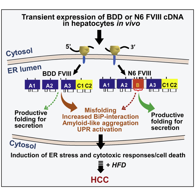
Hemophilia A gene therapy targets hepatocytes to express B domain deleted (BDD) clotting factor VIII (FVIII) to permit viral encapsidation. Since BDD is prone to misfolding in the endoplasmic reticulum (ER) and ER protein misfolding in hepatocytes followed by high-fat diet (HFD) can cause hepatocellular carcinoma (HCC), we studied how FVIII misfolding impacts HCC development using hepatocyte DNA delivery to express three proteins from the same parental vector: (1) well-folded cytosolic dihydrofolate reductase (DHFR); (2) BDD-FVIII, which is prone to misfolding in the ER; and (3) N6-FVIII, which folds more efficiently than BDD-FVIII. One week after DNA delivery, when FVIII expression was undetectable, mice were fed HFD for 65 weeks. Remarkably, all mice that received BDD-FVIII vector developed liver tumors, whereas only 58% of mice that received N6 and no mice that received DHFR vector developed liver tumors, suggesting that the degree of protein misfolding in the ER increases predisposition to HCC in the context of an HFD and in the absence of viral transduction. Our findings raise concerns of ectopic BDD-FVIII expression in hepatocytes in the clinic, which poses risks independent of viral vector integration. Limited expression per hepatocyte and/or use of proteins that avoid misfolding may enhance safety.
血友病 A 基因治疗靶向肝细胞表达 B 结构域缺失(BDD)凝血因子 VIII(FVIII)以允许病毒包裹。由于 BDD 在内质网(ER)中容易错误折叠,并且 ER 蛋白在肝细胞中的错误折叠随后进行高脂肪饮食(HFD)可能导致肝细胞癌(HCC),我们研究了 FVIII 错误折叠如何影响 HCC 的发展,方法是使用肝细胞 DNA 传递来表达来自同一亲本载体的三种蛋白质:(1)折叠良好的细胞质二氢叶酸还原酶(DHFR);(2)BDD-FVIII,其在 ER 中容易错误折叠;和(3)N6-FVIII,其折叠效率高于 BDD-FVIII。在 DNA 传递后一周,当 FVIII 表达无法检测到时,用 HFD 喂养小鼠 65 周。值得注意的是,接受 BDD-FVIII 载体的所有小鼠均发展为肝肿瘤,而仅接受 N6 的 58%的小鼠和未接受 DHFR 载体的小鼠未发展为肝肿瘤,表明 ER 中的蛋白错误折叠程度增加了在 HFD 存在和没有病毒转导的情况下 HCC 的易感性。我们的发现引起了对临床中肝细胞中异位 BDD-FVIII 表达的担忧,这独立于病毒载体整合带来风险。每个肝细胞的表达受限和/或使用避免错误折叠的蛋白质可能会提高安全性。