Respiratory Medicine, The University of Hong Kong-Shenzhen Hospital, Shenzhen, China.
Respiratory Medicine, The University of Hong Kong-Shenzhen Hospital, Shenzhen, China; Department of Medicine, The University of Hong Kong, Hong Kong SAR, China.
Biomed J. 2023 Oct;46(5):100566. doi: 10.1016/j.bj.2022.10.003. Epub 2022 Oct 13.
Both obstructive sleep apnea (OSA) and non-alcoholic fatty liver disease (NAFLD) are prevalent within obese individuals. We aimed to investigate the effects of intermittent hypoxia (IH), a clinical feature of OSA, on hepatic expression of fatty acid translocase (CD36) in relation to liver injury in lean and diet-induced obese mice.
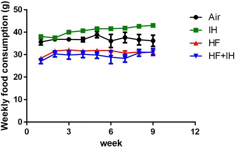
Four-week-old male C57BL/6J mice were randomized to standard diet (SD) or high fat (HF) diet groups. At 13-week-old, all mice were exposed to either air or IH (IH30; thirty hypoxic episodes per hour) for four weeks. We assessed liver injury through lipid profile, oxidative and inflammatory stress, histological scoring and hepatic CD36 expression.
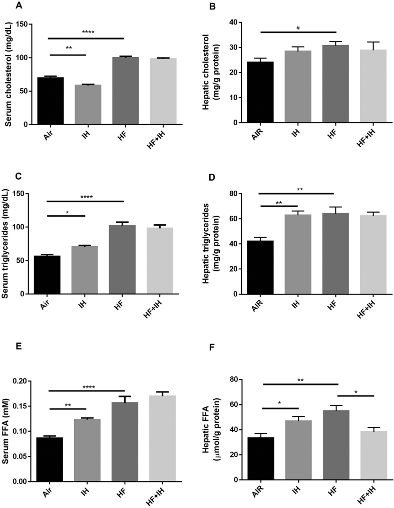
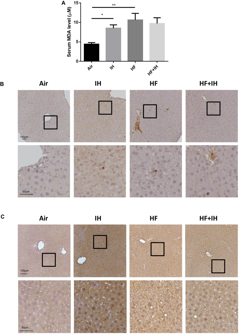
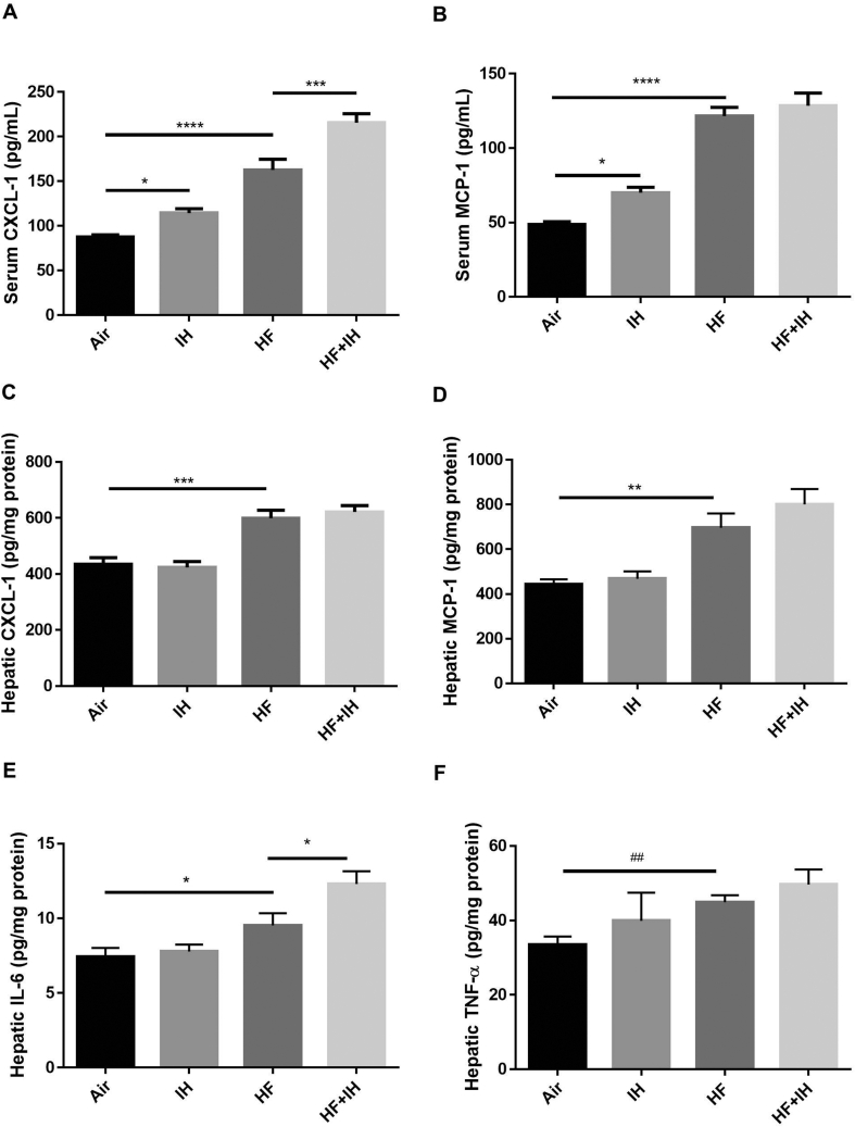
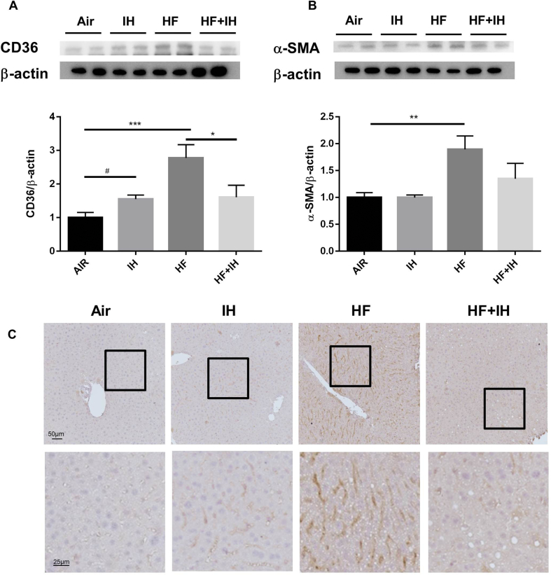
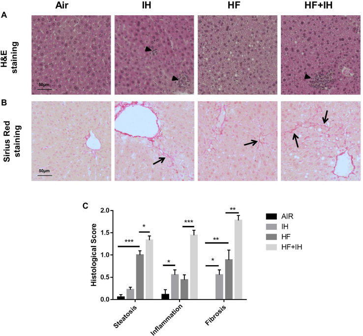
In lean mice, IH elevated serum and hepatic triglyceride and free fatty acid (FFA) levels, in line with upregulation of hepatic CD36 expression and myeloperoxidase (MPO)-positive cells in support of inflammatory infiltrates along with increase in serum malondialdehyde (MDA), C-X-C motif chemokine ligand 1(CXCL-1) and monocyte chemoattractant protein-1 (MCP-1). In diet-induced obese mice, an increase in hepatic alanine transaminase (ALT) activity, serum and hepatic levels of lipid parameters and inflammatory markers, serum MDA level, hepatic expressions of CD36 and α-smooth muscle actin (α-SMA), and MPO-positive cells was observed. IH potentiated hepatic ALT activity, serum CXCL-1 and hepatic interleukin-6 (IL-6), in line with inflammatory infiltrates, but paradoxically, reduced hepatic FFA level and hepatic CD36 expression, compared to obese mice without IH exposure. However, IH further augmented diet-induced liver steatosis and fibrosis as shown by histological scores.
This study contributes to support that IH featuring OSA may lead to liver injury via differential regulation of hepatic CD36 expression in lean and diet-induced obese mice.
阻塞性睡眠呼吸暂停(OSA)和非酒精性脂肪性肝病(NAFLD)在肥胖人群中都很常见。我们旨在研究间歇性低氧(IH),即 OSA 的一个临床特征,对瘦鼠和饮食诱导肥胖鼠肝内脂肪酸转位酶(CD36)表达与肝损伤的影响。
将 4 周龄雄性 C57BL/6J 小鼠随机分为标准饮食(SD)或高脂肪(HF)饮食组。13 周龄时,所有小鼠均暴露于空气或 IH(IH30;每小时 30 次缺氧发作)4 周。我们通过血脂谱、氧化和炎症应激、组织学评分和肝 CD36 表达来评估肝损伤。
在瘦鼠中,IH 升高了血清和肝甘油三酯和游离脂肪酸(FFA)水平,与肝 CD36 表达上调和髓过氧化物酶(MPO)阳性细胞一致,支持炎症浸润,并伴有血清丙二醛(MDA)、C-X-C 基序趋化因子配体 1(CXCL-1)和单核细胞趋化蛋白-1(MCP-1)增加。在饮食诱导肥胖鼠中,观察到肝丙氨酸氨基转移酶(ALT)活性、血清和肝脂质参数和炎症标志物、血清 MDA 水平、肝 CD36 和α-平滑肌肌动蛋白(α-SMA)表达以及 MPO 阳性细胞增加。与未暴露于 IH 的肥胖小鼠相比,IH 增加了肝 ALT 活性、血清 CXCL-1 和肝白细胞介素-6(IL-6),并伴有炎症浸润,但出人意料的是,降低了肝 FFA 水平和肝 CD36 表达。然而,与未暴露于 IH 的肥胖小鼠相比,IH 进一步加重了饮食诱导的肝脂肪变性和纤维化,这可以通过组织学评分来证明。
这项研究支持 OSA 特征的 IH 通过在瘦鼠和饮食诱导肥胖鼠中对肝 CD36 表达的差异调节导致肝损伤。