• 文献检索
  • 文档翻译
  • 深度研究
  • 学术资讯
  • Suppr Zotero 插件Zotero 插件
  • 邀请有礼
  • 套餐&价格
  • 历史记录
应用&插件
Suppr Zotero 插件Zotero 插件浏览器插件Mac 客户端Windows 客户端微信小程序
定价
高级版会员购买积分包购买API积分包
服务
文献检索文档翻译深度研究API 文档MCP 服务
关于我们
关于 Suppr公司介绍联系我们用户协议隐私条款
关注我们

Suppr 超能文献

核心技术专利:CN118964589B侵权必究
粤ICP备2023148730 号-1Suppr @ 2026

文献检索

告别复杂PubMed语法,用中文像聊天一样搜索,搜遍4000万医学文献。AI智能推荐,让科研检索更轻松。

立即免费搜索

文件翻译

保留排版,准确专业,支持PDF/Word/PPT等文件格式,支持 12+语言互译。

免费翻译文档

深度研究

AI帮你快速写综述,25分钟生成高质量综述,智能提取关键信息,辅助科研写作。

立即免费体验

C3G 的下调通过促进上皮-间充质样过程增强卵圆细胞的促迁移和干性特性。

C3G down-regulation enhances pro-migratory and stemness properties of oval cells by promoting an epithelial-mesenchymal-like process.

机构信息

Departamento de Bioquímica y Biología Molecular, Facultad de Farmacia, Universidad Complutense de Madrid; 28040 Madrid, Spain.

Instituto de Investigación Sanitaria del Hospital Clínico San Carlos (IdISSC), 28040 Madrid, Spain.

出版信息

Int J Biol Sci. 2022 Sep 25;18(15):5873-5884. doi: 10.7150/ijbs.73192. eCollection 2022.

DOI:10.7150/ijbs.73192
PMID:36263169
原文链接:https://pmc.ncbi.nlm.nih.gov/articles/PMC9576514/
Abstract

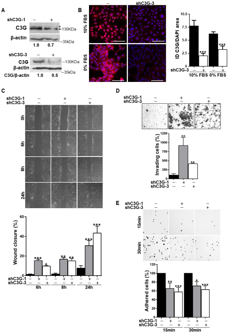
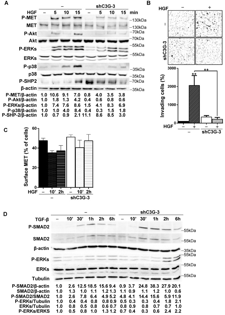
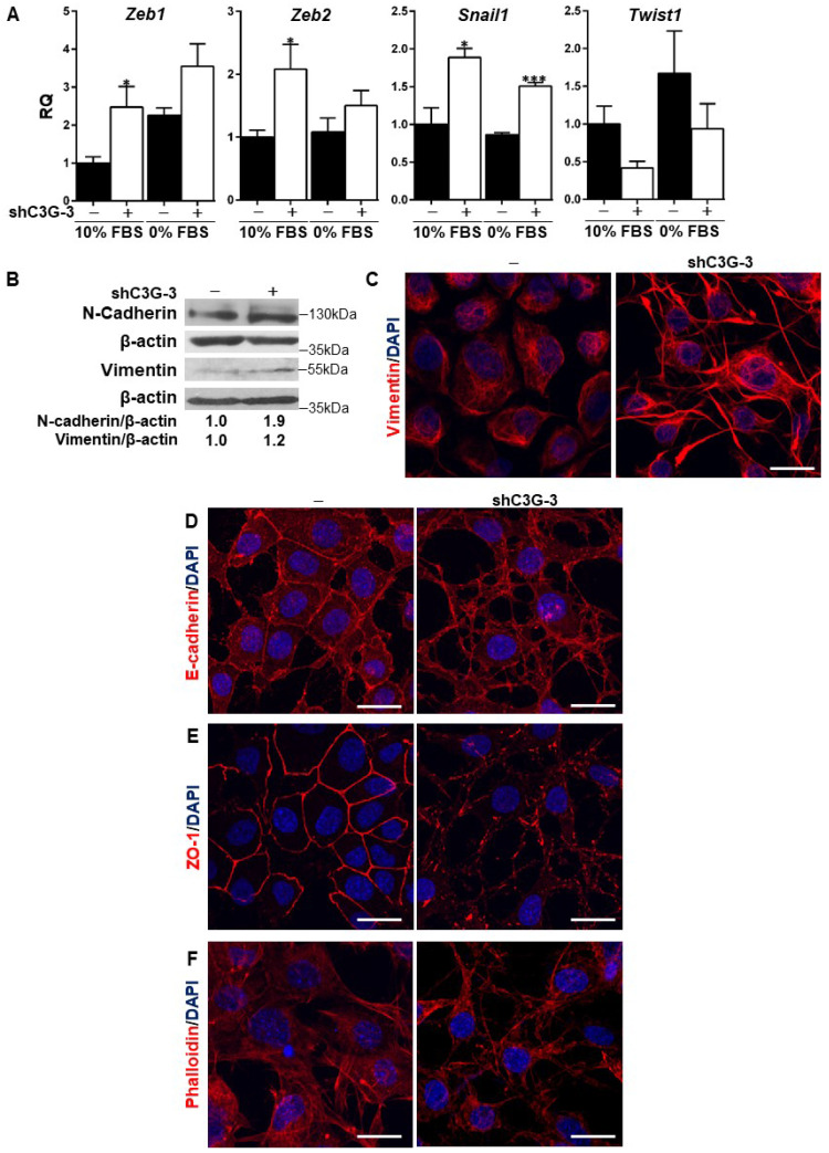
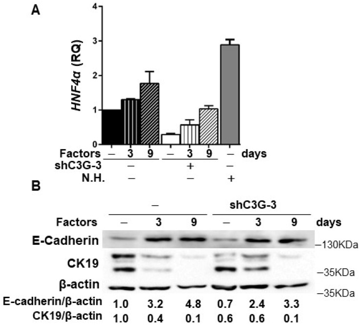
Previous data indicate that C3G (RapGEF1) main isoform is highly expressed in liver progenitor cells (or oval cells) compared to adult mature hepatocytes, suggesting it may play an important role in oval cell biology. Hence, we have explored C3G function in the regulation of oval cell properties by permanent gene silencing using shRNAs. We found that C3G knock-down enhanced migratory and invasive ability of oval cells by promoting a partial epithelial to mesenchymal transition (EMT). This is likely mediated by upregulation of mRNA expression of the EMT-inducing transcription factors, , and , induced in C3G-silenced oval cells. This EMT is associated to a higher expression of the stemness markers, CD133 and CD44. Moreover, C3G down-regulation increased oval cells clonogenic capacity by enhancing cell scattering. However, C3G knock-down did not impair oval cell differentiation into hepatocyte lineage. Mechanistic studies revealed that HGF/MET signaling and its pro-invasive activity was impaired in oval cells with low levels of C3G, while TGF-β signaling was increased. Altogether, these data suggest that C3G might be tightly regulated to ensure liver repair in chronic liver diseases such as non-alcoholic steatohepatitis. Hence, reduced C3G levels could facilitate oval cell expansion, after the proliferation peak, by enhancing migration.

摘要

先前的数据表明,与成年成熟肝细胞相比,C3G(RapGEF1)的主要同工型在肝祖细胞(或卵圆细胞)中高度表达,这表明它可能在卵圆细胞生物学中发挥重要作用。因此,我们通过使用 shRNA 进行永久基因沉默来探索 C3G 在调节卵圆细胞特性方面的功能。我们发现,C3G 敲低通过促进部分上皮间质转化(EMT)来增强卵圆细胞的迁移和侵袭能力。这可能是通过 C3G 沉默的卵圆细胞中 EMT 诱导转录因子、、和的 mRNA 表达上调介导的。这种 EMT 与干细胞标志物 CD133 和 CD44 的高表达相关。此外,C3G 下调通过增强细胞散射增加了卵圆细胞的集落形成能力。然而,C3G 敲低并没有损害卵圆细胞向肝谱系的分化。机制研究表明,在 C3G 水平较低的卵圆细胞中,HGF/MET 信号及其促侵袭活性受损,而 TGF-β 信号增强。总的来说,这些数据表明,C3G 可能受到严格调控,以确保慢性肝病(如非酒精性脂肪性肝炎)中的肝脏修复。因此,C3G 水平降低可能会通过增强迁移来促进增殖高峰期后卵圆细胞的扩张。

https://cdn.ncbi.nlm.nih.gov/pmc/blobs/7532/9576514/bdb3685656d6/ijbsv18p5873g005.jpg
https://cdn.ncbi.nlm.nih.gov/pmc/blobs/7532/9576514/603f34c536a1/ijbsv18p5873g001.jpg
https://cdn.ncbi.nlm.nih.gov/pmc/blobs/7532/9576514/5f735072aa1a/ijbsv18p5873g002.jpg
https://cdn.ncbi.nlm.nih.gov/pmc/blobs/7532/9576514/5a1335046267/ijbsv18p5873g003.jpg
https://cdn.ncbi.nlm.nih.gov/pmc/blobs/7532/9576514/07038c1ebecc/ijbsv18p5873g004.jpg
https://cdn.ncbi.nlm.nih.gov/pmc/blobs/7532/9576514/bdb3685656d6/ijbsv18p5873g005.jpg

相似文献

1
C3G down-regulation enhances pro-migratory and stemness properties of oval cells by promoting an epithelial-mesenchymal-like process.C3G 的下调通过促进上皮-间充质样过程增强卵圆细胞的促迁移和干性特性。
Int J Biol Sci. 2022 Sep 25;18(15):5873-5884. doi: 10.7150/ijbs.73192. eCollection 2022.
2
c-Met Signaling Is Essential for Mouse Adult Liver Progenitor Cells Expansion After Transforming Growth Factor-β-Induced Epithelial-Mesenchymal Transition and Regulates Cell Phenotypic Switch.c-Met 信号对转化生长因子-β诱导的上皮-间充质转化后小鼠成体肝祖细胞的扩增至关重要,并调节细胞表型转换。
Stem Cells. 2019 Aug;37(8):1108-1118. doi: 10.1002/stem.3038. Epub 2019 Jun 18.
3
C3G Is Upregulated in Hepatocarcinoma, Contributing to Tumor Growth and Progression and to HGF/MET Pathway Activation.补体C3肾小球肾炎因子在肝癌中上调,促进肿瘤生长、进展以及肝细胞生长因子/间质-上皮转化因子通路激活。
Cancers (Basel). 2020 Aug 14;12(8):2282. doi: 10.3390/cancers12082282.
4
Transforming growth factor-β-induced plasticity causes a migratory stemness phenotype in hepatocellular carcinoma.转化生长因子-β诱导的可塑性导致肝细胞癌出现迁移性干性表型。
Cancer Lett. 2017 Apr 28;392:39-50. doi: 10.1016/j.canlet.2017.01.037. Epub 2017 Feb 2.
5
HGF/c-Met signaling promotes liver progenitor cell migration and invasion by an epithelial-mesenchymal transition-independent, phosphatidyl inositol-3 kinase-dependent pathway in an in vitro model.在体外模型中,HGF/c-Met信号通过一条不依赖上皮-间质转化、依赖磷脂酰肌醇-3激酶的途径促进肝祖细胞的迁移和侵袭。
Biochim Biophys Acta. 2015 Oct;1853(10 Pt A):2453-63. doi: 10.1016/j.bbamcr.2015.05.017. Epub 2015 May 20.
6
Expression of extracellular matrix genes in cultured hepatic oval cells: an origin of hepatic stellate cells through transforming growth factor beta?培养的肝卵圆细胞中细胞外基质基因的表达:通过转化生长因子β产生肝星状细胞的一个来源?
Liver Int. 2009 Apr;29(4):575-84. doi: 10.1111/j.1478-3231.2009.01992.x.
7
OVOL2 links stemness and metastasis via fine-tuning epithelial-mesenchymal transition in nasopharyngeal carcinoma.OVOL2 通过精细调控鼻咽癌中的上皮-间充质转化来连接干性和转移。
Theranostics. 2018 Mar 8;8(8):2202-2216. doi: 10.7150/thno.24003. eCollection 2018.
8
Epithelial-mesenchymal transition transcription factor ZEB1/ZEB2 co-expression predicts poor prognosis and maintains tumor-initiating properties in head and neck cancer.上皮-间充质转化转录因子 ZEB1/ZEB2 共表达预测头颈部癌症预后不良,并维持肿瘤起始特性。
Oral Oncol. 2013 Jan;49(1):34-41. doi: 10.1016/j.oraloncology.2012.07.012. Epub 2012 Aug 11.
9
Down-regulation of FXYD3 is induced by transforming growth factor-β signaling via ZEB1/δEF1 in human mammary epithelial cells.转化生长因子-β信号通过 ZEB1/δEF1 诱导人乳腺上皮细胞中 FXYD3 的下调。
Biol Pharm Bull. 2011;34(3):324-9. doi: 10.1248/bpb.34.324.
10
Berberine induces neuronal differentiation through inhibition of cancer stemness and epithelial-mesenchymal transition in neuroblastoma cells.黄连素通过抑制神经母细胞瘤细胞的癌干性和上皮-间质转化诱导神经元分化。
Phytomedicine. 2016 Jun 15;23(7):736-44. doi: 10.1016/j.phymed.2016.03.013. Epub 2016 Apr 13.

引用本文的文献

1
C3G promotes bone marrow adipocyte expansion and hematopoietic regeneration after myeloablation by enhancing megakaryocyte niche function.C3G通过增强巨核细胞龛功能促进骨髓消融后骨髓脂肪细胞扩张和造血再生。
J Hematol Oncol. 2025 Apr 1;18(1):38. doi: 10.1186/s13045-025-01687-1.
2
Platelet C3G protects from liver fibrosis, while enhancing tumor growth through regulation of the immune response.血小板C3G可预防肝纤维化,同时通过调节免疫反应促进肿瘤生长。
J Pathol. 2025 Apr;265(4):502-517. doi: 10.1002/path.6403. Epub 2025 Feb 24.
3
Cepharanthine inhibits migration, invasion, and EMT of bladder cancer cells by activating the Rap1 signaling pathway in vitro.

本文引用的文献

1
C3G Protein, a New Player in Glioblastoma.C3G 蛋白,胶质母细胞瘤的新角色。
Int J Mol Sci. 2021 Sep 16;22(18):10018. doi: 10.3390/ijms221810018.
2
C3G downregulation induces the acquisition of a mesenchymal phenotype that enhances aggressiveness of glioblastoma cells.C3G 的下调诱导获得间充质表型,从而增强神经胶质瘤细胞的侵袭性。
Cell Death Dis. 2021 Apr 6;12(4):348. doi: 10.1038/s41419-021-03631-w.
3
C3G Regulates STAT3, ERK, Adhesion Signaling, and Is Essential for Differentiation of Embryonic Stem Cells.C3G 调节 STAT3、ERK、黏附信号,并对胚胎干细胞的分化至关重要。
千金藤素在体外通过激活Rap1信号通路抑制膀胱癌细胞的迁移、侵袭和上皮-间质转化。
Am J Transl Res. 2024 May 15;16(5):1602-1619. doi: 10.62347/WDFF7432. eCollection 2024.
4
New and Old Key Players in Liver Cancer.肝癌的新老关键角色。
Int J Mol Sci. 2023 Dec 5;24(24):17152. doi: 10.3390/ijms242417152.
Stem Cell Rev Rep. 2021 Aug;17(4):1465-1477. doi: 10.1007/s12015-021-10136-8. Epub 2021 Feb 23.
4
Expression of a novel brain specific isoform of C3G is regulated during development.一种新型脑特异性 C3G 同工型的表达在发育过程中受到调控。
Sci Rep. 2020 Nov 2;10(1):18838. doi: 10.1038/s41598-020-75813-z.
5
C3G Is Upregulated in Hepatocarcinoma, Contributing to Tumor Growth and Progression and to HGF/MET Pathway Activation.补体C3肾小球肾炎因子在肝癌中上调,促进肿瘤生长、进展以及肝细胞生长因子/间质-上皮转化因子通路激活。
Cancers (Basel). 2020 Aug 14;12(8):2282. doi: 10.3390/cancers12082282.
6
EpCAM inhibits differentiation of human liver progenitor cells into hepatocytes in vitro by activating Notch1 signaling.上皮细胞黏附分子(EpCAM)通过激活Notch1信号通路在体外抑制人肝祖细胞向肝细胞的分化。
Biochem Biophys Res Commun. 2020 Feb 19. doi: 10.1016/j.bbrc.2020.02.041.
7
Liver Progenitors and Adult Cell Plasticity in Hepatic Injury and Repair: Knowns and Unknowns.肝祖细胞与成体肝干细胞在肝损伤与修复中的可塑性:已知与未知。
Annu Rev Pathol. 2020 Jan 24;15:23-50. doi: 10.1146/annurev-pathmechdis-012419-032824. Epub 2019 Aug 9.
8
c-Met Signaling Is Essential for Mouse Adult Liver Progenitor Cells Expansion After Transforming Growth Factor-β-Induced Epithelial-Mesenchymal Transition and Regulates Cell Phenotypic Switch.c-Met 信号对转化生长因子-β诱导的上皮-间充质转化后小鼠成体肝祖细胞的扩增至关重要,并调节细胞表型转换。
Stem Cells. 2019 Aug;37(8):1108-1118. doi: 10.1002/stem.3038. Epub 2019 Jun 18.
9
C3G, through its GEF activity, induces megakaryocytic differentiation and proplatelet formation.C3G 通过其 GEF 活性诱导巨核细胞分化和血小板形成。
Cell Commun Signal. 2018 Dec 19;16(1):101. doi: 10.1186/s12964-018-0311-5.
10
How Rap and its GEFs control liver physiology and cancer development. C3G alterations in human hepatocarcinoma.说唱乐及其鸟嘌呤核苷酸交换因子如何控制肝脏生理和癌症发展。人类肝癌中的C3G改变。
Hepat Oncol. 2018 Apr 16;5(1):HEP05. doi: 10.2217/hep-2017-0026. eCollection 2018 Jan.