• 文献检索
  • 文档翻译
  • 深度研究
  • 学术资讯
  • Suppr Zotero 插件Zotero 插件
  • 邀请有礼
  • 套餐&价格
  • 历史记录
应用&插件
Suppr Zotero 插件Zotero 插件浏览器插件Mac 客户端Windows 客户端微信小程序
定价
高级版会员购买积分包购买API积分包
服务
文献检索文档翻译深度研究API 文档MCP 服务
关于我们
关于 Suppr公司介绍联系我们用户协议隐私条款
关注我们

Suppr 超能文献

核心技术专利:CN118964589B侵权必究
粤ICP备2023148730 号-1Suppr @ 2026

文献检索

告别复杂PubMed语法,用中文像聊天一样搜索,搜遍4000万医学文献。AI智能推荐,让科研检索更轻松。

立即免费搜索

文件翻译

保留排版,准确专业,支持PDF/Word/PPT等文件格式,支持 12+语言互译。

免费翻译文档

深度研究

AI帮你快速写综述,25分钟生成高质量综述,智能提取关键信息,辅助科研写作。

立即免费体验

补体C3肾小球肾炎因子在肝癌中上调,促进肿瘤生长、进展以及肝细胞生长因子/间质-上皮转化因子通路激活。

C3G Is Upregulated in Hepatocarcinoma, Contributing to Tumor Growth and Progression and to HGF/MET Pathway Activation.

作者信息

Sequera Celia, Bragado Paloma, Manzano Sara, Arechederra Maria, Richelme Sylvie, Gutiérrez-Uzquiza Alvaro, Sánchez Aránzazu, Maina Flavio, Guerrero Carmen, Porras Almudena

机构信息

Departamento de Bioquímica y Biología Molecular, Facultad de Farmacia, Universidad Complutense de Madrid, 28040 Madrid, Spain.

Instituto de Investigación Sanitaria del Hospital Clínico San Carlos (IdISSC), 28040 Madrid, Spain.

出版信息

Cancers (Basel). 2020 Aug 14;12(8):2282. doi: 10.3390/cancers12082282.

DOI:10.3390/cancers12082282
PMID:32823931
原文链接:https://pmc.ncbi.nlm.nih.gov/articles/PMC7463771/
Abstract

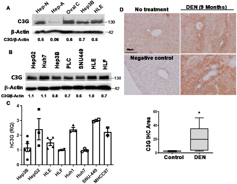
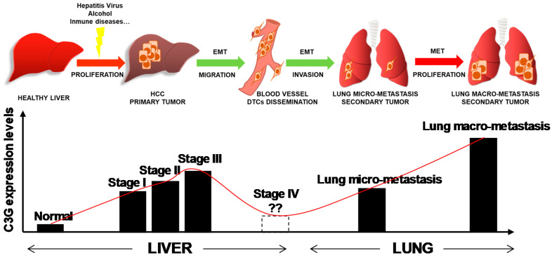
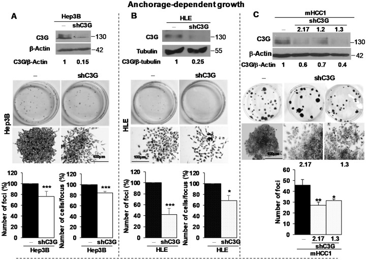
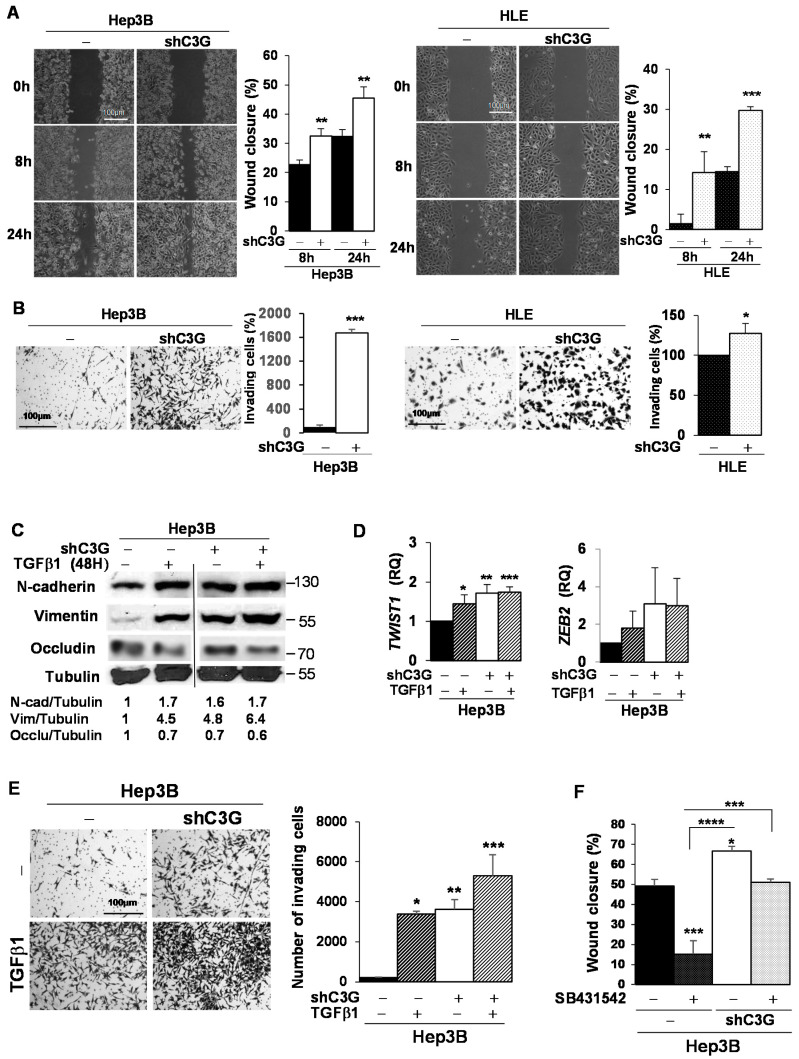
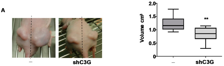
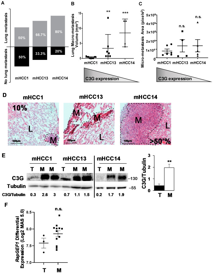
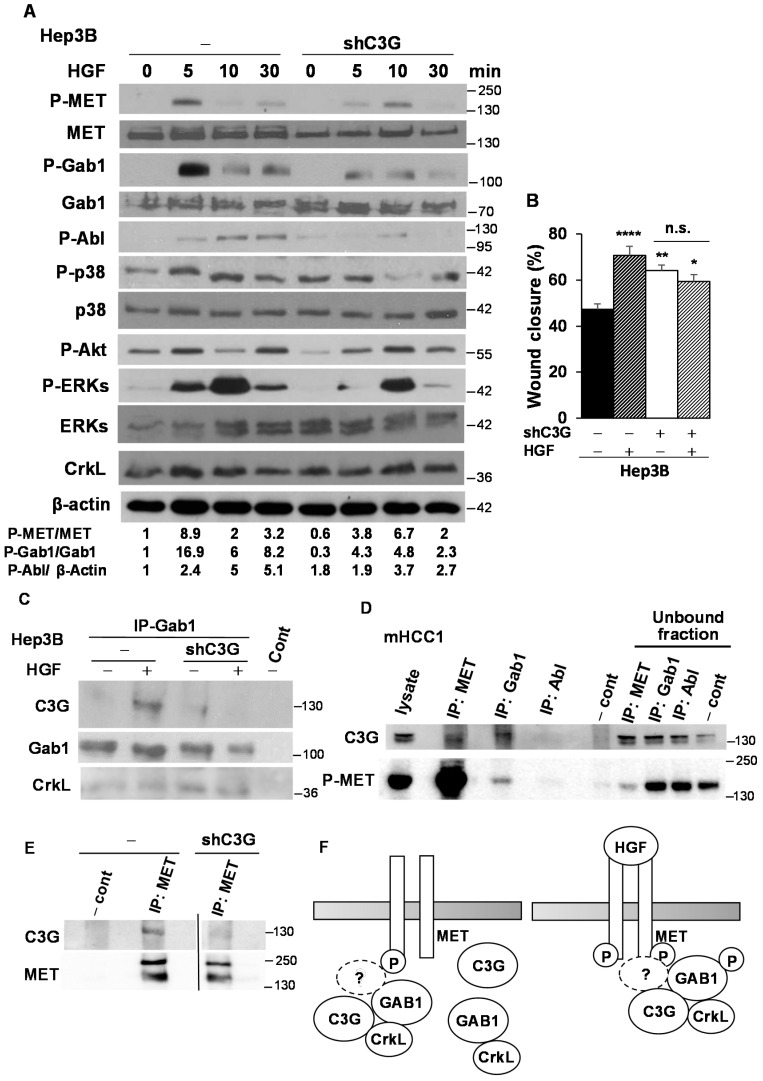
The complexity of hepatocellular carcinoma (HCC) challenges the identification of disease-relevant signals. C3G, a guanine nucleotide exchange factor for Rap and other Ras proteins, plays a dual role in cancer acting as either a tumor suppressor or promoter depending on tumor type and stage. The potential relevance of C3G upregulation in HCC patients suggested by database analysis remains unknown. We have explored C3G function in HCC and the underlying mechanisms using public patient data and in vitro and in vivo human and mouse HCC models. We found that C3G is highly expressed in progenitor cells and neonatal hepatocytes, whilst being down-regulated in adult hepatocytes and re-expressed in human HCC patients, mouse HCC models and HCC cell lines. Moreover, high mRNA levels correlate with tumor progression and a lower patient survival rate. C3G expression appears to be tightly modulated within the HCC program, influencing distinct cell biological properties. Hence, high C3G expression levels are necessary for cell tumorigenic properties, as illustrated by reduced colony formation in anchorage-dependent and -independent growth assays induced by permanent C3G silencing using shRNAs. Additionally, we demonstrate that C3G down-regulation interferes with primary HCC tumor formation in xenograft assays, increasing apoptosis and decreasing proliferation. In vitro assays also revealed that C3G down-regulation enhances the pro-migratory, invasive and metastatic properties of HCC cells through an epithelial-mesenchymal switch that favors the acquisition of a more mesenchymal phenotype. Consistently, a low C3G expression in HCC cells correlates with lung metastasis formation in mice. However, the subsequent restoration of C3G levels is associated with metastatic growth. Mechanistically, C3G down-regulation severely impairs HGF/MET signaling activation in HCC cells. Collectively, our results indicate that C3G is a key player in HCC. C3G promotes tumor growth and progression, and the modulation of its levels is essential to ensure distinct biological features of HCC cells throughout the oncogenic program. Furthermore, C3G requirement for HGF/MET signaling full activation provides mechanistic data on how it works, pointing out the relevance of assessing whether high C3G levels could identify HCC responders to MET inhibitors.

摘要

肝细胞癌(HCC)的复杂性对疾病相关信号的识别提出了挑战。C3G是一种针对Rap和其他Ras蛋白的鸟嘌呤核苷酸交换因子,在癌症中发挥双重作用,根据肿瘤类型和阶段,既可以作为肿瘤抑制因子,也可以作为肿瘤促进因子。数据库分析提示的C3G上调在HCC患者中的潜在相关性尚不清楚。我们利用公开的患者数据以及体外和体内的人源和鼠源HCC模型,探索了C3G在HCC中的功能及其潜在机制。我们发现C3G在祖细胞和新生肝细胞中高表达,而在成年肝细胞中下调,并在人类HCC患者、小鼠HCC模型和HCC细胞系中重新表达。此外,高mRNA水平与肿瘤进展和较低的患者生存率相关。C3G的表达似乎在HCC进程中受到严格调控,影响着不同的细胞生物学特性。因此,高C3G表达水平是细胞致瘤特性所必需的,使用shRNA永久沉默C3G可导致贴壁依赖性和非依赖性生长试验中的集落形成减少,这证明了这一点。此外,我们证明在异种移植试验中,C3G下调会干扰原发性HCC肿瘤的形成,增加细胞凋亡并减少增殖。体外试验还表明,C3G下调通过上皮-间质转化增强HCC细胞的促迁移、侵袭和转移特性,这种转化有利于获得更具间质表型。一致的是,HCC细胞中低C3G表达与小鼠肺转移形成相关。然而,随后C3G水平的恢复与转移生长相关。从机制上讲,C3G下调会严重损害HCC细胞中HGF/MET信号的激活。总体而言,我们的结果表明C3G是HCC中的关键因子。C3G促进肿瘤生长和进展,其水平的调节对于确保HCC细胞在整个致癌过程中的独特生物学特征至关重要。此外,C3G对HGF/MET信号完全激活的需求提供了其作用机制的数据,指出了评估高C3G水平是否可识别对MET抑制剂有反应的HCC患者的相关性。

https://cdn.ncbi.nlm.nih.gov/pmc/blobs/269f/7463771/ecc261a4344f/cancers-12-02282-g007.jpg
https://cdn.ncbi.nlm.nih.gov/pmc/blobs/269f/7463771/f02759c5eb6c/cancers-12-02282-g001a.jpg
https://cdn.ncbi.nlm.nih.gov/pmc/blobs/269f/7463771/f20a0f424668/cancers-12-02282-g002a.jpg
https://cdn.ncbi.nlm.nih.gov/pmc/blobs/269f/7463771/fd1bfbd5b6a6/cancers-12-02282-g003.jpg
https://cdn.ncbi.nlm.nih.gov/pmc/blobs/269f/7463771/0e4954c1a4d2/cancers-12-02282-g004a.jpg
https://cdn.ncbi.nlm.nih.gov/pmc/blobs/269f/7463771/ec8601f40ecd/cancers-12-02282-g005.jpg
https://cdn.ncbi.nlm.nih.gov/pmc/blobs/269f/7463771/3aa85e5523ae/cancers-12-02282-g006.jpg
https://cdn.ncbi.nlm.nih.gov/pmc/blobs/269f/7463771/ecc261a4344f/cancers-12-02282-g007.jpg

相似文献

1
C3G Is Upregulated in Hepatocarcinoma, Contributing to Tumor Growth and Progression and to HGF/MET Pathway Activation.补体C3肾小球肾炎因子在肝癌中上调,促进肿瘤生长、进展以及肝细胞生长因子/间质-上皮转化因子通路激活。
Cancers (Basel). 2020 Aug 14;12(8):2282. doi: 10.3390/cancers12082282.
2
C3G downregulation induces the acquisition of a mesenchymal phenotype that enhances aggressiveness of glioblastoma cells.C3G 的下调诱导获得间充质表型,从而增强神经胶质瘤细胞的侵袭性。
Cell Death Dis. 2021 Apr 6;12(4):348. doi: 10.1038/s41419-021-03631-w.
3
C3G down-regulation enhances pro-migratory and stemness properties of oval cells by promoting an epithelial-mesenchymal-like process.C3G 的下调通过促进上皮-间充质样过程增强卵圆细胞的促迁移和干性特性。
Int J Biol Sci. 2022 Sep 25;18(15):5873-5884. doi: 10.7150/ijbs.73192. eCollection 2022.
4
C3G knock-down enhances migration and invasion by increasing Rap1-mediated p38α activation, while it impairs tumor growth through p38α-independent mechanisms.C3G基因敲低通过增加Rap1介导的p38α激活来增强迁移和侵袭能力,而它通过不依赖p38α的机制损害肿瘤生长。
Oncotarget. 2016 Jul 19;7(29):45060-45078. doi: 10.18632/oncotarget.9911.
5
Attenuation of MET-mediated migration and invasion in hepatocellular carcinoma cells by SOCS1.SOCS1 抑制肝癌细胞中 MET 介导的迁移和侵袭。
World J Gastroenterol. 2017 Sep 28;23(36):6639-6649. doi: 10.3748/wjg.v23.i36.6639.
6
How Rap and its GEFs control liver physiology and cancer development. C3G alterations in human hepatocarcinoma.说唱乐及其鸟嘌呤核苷酸交换因子如何控制肝脏生理和癌症发展。人类肝癌中的C3G改变。
Hepat Oncol. 2018 Apr 16;5(1):HEP05. doi: 10.2217/hep-2017-0026. eCollection 2018 Jan.
7
Regulation of MET receptor tyrosine kinase signaling by suppressor of cytokine signaling 1 in hepatocellular carcinoma.细胞因子信号转导抑制因子1对肝细胞癌中MET受体酪氨酸激酶信号通路的调控
Oncogene. 2015 Nov 12;34(46):5718-28. doi: 10.1038/onc.2015.20. Epub 2015 Mar 2.
8
Induction of tumor initiation is dependent on CD44s in c-Met⁺ hepatocellular carcinoma.在c-Met⁺肝细胞癌中,肿瘤起始的诱导依赖于CD44s。
BMC Cancer. 2015 Mar 21;15:161. doi: 10.1186/s12885-015-1166-4.
9
C3G down-regulates p38 MAPK activity in response to stress by Rap-1 independent mechanisms: involvement in cell death.C3G 通过独立于 Rap-1 的机制来下调应激时的 p38 MAPK 活性:参与细胞死亡。
Cell Signal. 2010 Mar;22(3):533-42. doi: 10.1016/j.cellsig.2009.11.008.
10
Regulator of Calcineurin 1 Gene Isoform 4, Down-regulated in Hepatocellular Carcinoma, Prevents Proliferation, Migration, and Invasive Activity of Cancer Cells and Metastasis of Orthotopic Tumors by Inhibiting Nuclear Translocation of NFAT1.钙调神经磷酸酶 1 基因同种型 4 调节物,在肝癌中下调,通过抑制 NFAT1 的核易位来防止癌细胞的增殖、迁移和侵袭活性以及原位肿瘤的转移。
Gastroenterology. 2017 Sep;153(3):799-811.e33. doi: 10.1053/j.gastro.2017.05.045. Epub 2017 Jun 2.

引用本文的文献

1
Platelet C3G protects from liver fibrosis, while enhancing tumor growth through regulation of the immune response.血小板C3G可预防肝纤维化,同时通过调节免疫反应促进肿瘤生长。
J Pathol. 2025 Apr;265(4):502-517. doi: 10.1002/path.6403. Epub 2025 Feb 24.
2
New and Old Key Players in Liver Cancer.肝癌的新老关键角色。
Int J Mol Sci. 2023 Dec 5;24(24):17152. doi: 10.3390/ijms242417152.
3
Bioinformatics analysis and experimental validation of a novel autophagy-related signature relevant to immune infiltration for recurrence prediction after curative hepatectomy.

本文引用的文献

1
Deficiency in Embryonic Stem Cell Marker Reduced Expression 1 Activates Mitogen-Activated Protein Kinase Kinase 6-Dependent p38 Mitogen-Activated Protein Kinase Signaling to Drive Hepatocarcinogenesis.胚胎干细胞标志物缺失降低表达 1 激活丝裂原活化蛋白激酶激酶 6 依赖性 p38 丝裂原活化蛋白激酶信号通路,驱动肝癌发生。
Hepatology. 2020 Jul;72(1):183-197. doi: 10.1002/hep.31020. Epub 2020 Apr 8.
2
Tracking Dynamics of Spontaneous Tumors in Mice Using Photon-Counting Computed Tomography.使用光子计数计算机断层扫描技术追踪小鼠自发肿瘤的动态变化
iScience. 2019 Nov 22;21:68-83. doi: 10.1016/j.isci.2019.10.015. Epub 2019 Oct 9.
3
生物信息学分析和实验验证与免疫浸润相关的新型自噬相关特征,用于预测根治性肝切除术后的复发。
Aging (Albany NY). 2023 Apr 3;15(7):2610-2630. doi: 10.18632/aging.204632.
4
MYC and MET cooperatively drive hepatocellular carcinoma with distinct molecular traits and vulnerabilities.MYC 和 MET 协同驱动具有不同分子特征和脆弱性的肝细胞癌。
Cell Death Dis. 2022 Nov 24;13(11):994. doi: 10.1038/s41419-022-05411-6.
5
C3G down-regulation enhances pro-migratory and stemness properties of oval cells by promoting an epithelial-mesenchymal-like process.C3G 的下调通过促进上皮-间充质样过程增强卵圆细胞的促迁移和干性特性。
Int J Biol Sci. 2022 Sep 25;18(15):5873-5884. doi: 10.7150/ijbs.73192. eCollection 2022.
6
C3G Protein, a New Player in Glioblastoma.C3G 蛋白,胶质母细胞瘤的新角色。
Int J Mol Sci. 2021 Sep 16;22(18):10018. doi: 10.3390/ijms221810018.
7
C3G downregulation induces the acquisition of a mesenchymal phenotype that enhances aggressiveness of glioblastoma cells.C3G 的下调诱导获得间充质表型,从而增强神经胶质瘤细胞的侵袭性。
Cell Death Dis. 2021 Apr 6;12(4):348. doi: 10.1038/s41419-021-03631-w.
GREIN: An Interactive Web Platform for Re-analyzing GEO RNA-seq Data.
GREIN:一个用于重新分析 GEO RNA-seq 数据的交互式网络平台。
Sci Rep. 2019 May 20;9(1):7580. doi: 10.1038/s41598-019-43935-8.
4
C3G, through its GEF activity, induces megakaryocytic differentiation and proplatelet formation.C3G 通过其 GEF 活性诱导巨核细胞分化和血小板形成。
Cell Commun Signal. 2018 Dec 19;16(1):101. doi: 10.1186/s12964-018-0311-5.
5
Evaluating the landscape of gene cooperativity with receptor tyrosine kinases in liver tumorigenesis using transposon-mediated mutagenesis.利用转座子介导的诱变技术评估受体酪氨酸激酶在肝肿瘤发生中的基因协同作用景观。
J Hepatol. 2019 Mar;70(3):470-482. doi: 10.1016/j.jhep.2018.11.027. Epub 2018 Dec 6.
6
How Rap and its GEFs control liver physiology and cancer development. C3G alterations in human hepatocarcinoma.说唱乐及其鸟嘌呤核苷酸交换因子如何控制肝脏生理和癌症发展。人类肝癌中的C3G改变。
Hepat Oncol. 2018 Apr 16;5(1):HEP05. doi: 10.2217/hep-2017-0026. eCollection 2018 Jan.
7
Hypermethylation of gene body CpG islands predicts high dosage of functional oncogenes in liver cancer.基因体 CpG 岛的超甲基化预示着肝癌中功能性致癌基因的高剂量。
Nat Commun. 2018 Aug 8;9(1):3164. doi: 10.1038/s41467-018-05550-5.
8
Supervillin promotes epithelial-mesenchymal transition and metastasis of hepatocellular carcinoma in hypoxia via activation of the RhoA/ROCK-ERK/p38 pathway.Supervillin 通过激活 RhoA/ROCK-ERK/p38 通路促进低氧环境下肝癌的上皮-间充质转化和转移。
J Exp Clin Cancer Res. 2018 Jun 28;37(1):128. doi: 10.1186/s13046-018-0787-2.
9
Current and Future Treatment of Hepatocellular Carcinoma: An Updated Comprehensive Review.肝细胞癌的当前及未来治疗:最新全面综述
J Clin Transl Hepatol. 2018 Mar 28;6(1):69-78. doi: 10.14218/JCTH.2017.00031. Epub 2017 Dec 17.
10
Rap1b enhances the invasion and migration of hepatocellular carcinoma cells by up-regulating Twist 1.Rap1b 通过上调 Twist1 增强肝癌细胞的侵袭和迁移。
Exp Cell Res. 2018 Jun 1;367(1):56-64. doi: 10.1016/j.yexcr.2018.03.019. Epub 2018 Mar 17.