Departments of Nutrition and Movement Sciences, Maastricht, the Netherlands.
BioPharmaceuticals R&D, AstraZeneca, Gaithersburg, MD, USA.
Mol Metab. 2022 Dec;66:101620. doi: 10.1016/j.molmet.2022.101620. Epub 2022 Oct 21.
SGLT2 inhibitors increase urinary glucose excretion and have beneficial effects on cardiovascular and renal outcomes; the underlying mechanism may be metabolic adaptations due to urinary glucose loss. Here, we investigated the cellular and molecular effects of 5 weeks of dapagliflozin treatment on skeletal muscle metabolism in type 2 diabetes patients.
Twenty-six type 2 diabetes mellitus patients were randomized to a 5-week double-blind, cross-over study with 6-8-week wash-out. Skeletal muscle acetylcarnitine levels, intramyocellular lipid (IMCL) content and phosphocreatine (PCr) recovery rate were measured by magnetic resonance spectroscopy (MRS). Ex vivo mitochondrial respiration was measured in skeletal muscle fibers using high resolution respirometry. Intramyocellular lipid droplet and mitochondrial network dynamics were investigated using confocal microscopy. Skeletal muscle levels of acylcarnitines, amino acids and TCA cycle intermediates were measured. Expression of genes involved in fatty acid metabolism were investigated.
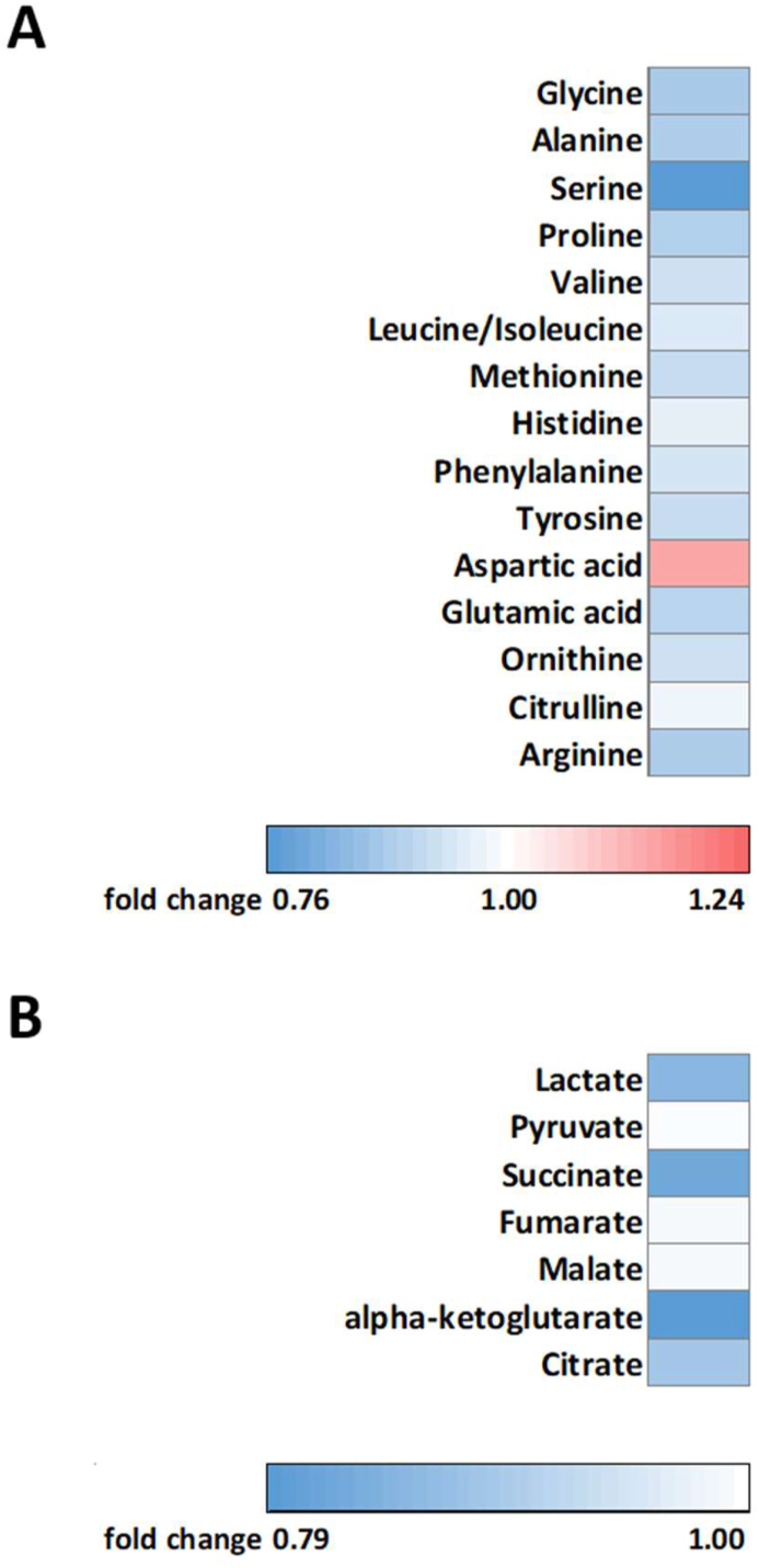
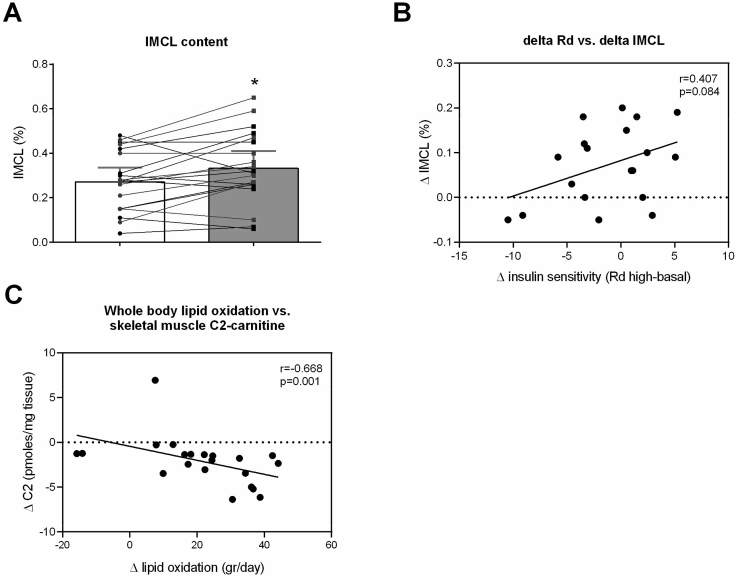
Mitochondrial function, mitochondrial network integrity and citrate synthase and carnitine acetyltransferase activities in skeletal muscle were unaltered after dapagliflozin treatment. Dapagliflozin treatment increased intramyocellular lipid content (0.060 (0.011, 0.110) %, p = 0.019). Myocellular lipid droplets increased in size (0.03 μm (0.01-0.06), p < 0.05) and number (0.003 μm (-0.001-0.007), p = 0.09) upon dapagliflozin treatment. CPT1A, CPT1B and malonyl CoA-decarboxylase mRNA expression was increased by dapagliflozin. Fasting acylcarnitine species and C4-OH carnitine levels (0.4704 (0.1246, 0.8162) pmoles∗mg tissue, p < 0.001) in skeletal muscle were higher after dapagliflozin treatment, while acetylcarnitine levels were lower (-40.0774 (-64.4766, -15.6782) pmoles∗mg tissue, p < 0.001). Fasting levels of several amino acids, succinate, alpha-ketoglutarate and lactate in skeletal muscle were significantly lower after dapagliflozin treatment.
Dapagliflozin treatment for 5 weeks leads to adaptive changes in skeletal muscle substrate metabolism favoring metabolism of fatty acid and ketone bodies and reduced glycolytic flux. The trial is registered with ClinicalTrials.gov, number NCT03338855.
SGLT2 抑制剂可增加尿糖排泄,并对心血管和肾脏结局有益;其潜在机制可能是由于尿糖丢失而导致的代谢适应。在这里,我们研究了 dapagliflozin 治疗 5 周对 2 型糖尿病患者骨骼肌代谢的细胞和分子影响。
26 名 2 型糖尿病患者被随机分配到为期 5 周的双盲交叉研究,6-8 周洗脱期。通过磁共振波谱(MRS)测量骨骼肌乙酰肉碱水平、细胞内脂质(IMCL)含量和磷酸肌酸(PCr)恢复率。使用高分辨率呼吸计测量骨骼肌纤维中的线粒体呼吸。使用共聚焦显微镜研究细胞内脂质滴和线粒体网络动力学。测量骨骼肌中酰基辅酶 A、氨基酸和 TCA 循环中间产物的水平。研究了与脂肪酸代谢相关的基因的表达。
dapagliflozin 治疗后,骨骼肌中的线粒体功能、线粒体网络完整性以及柠檬酸合酶和肉碱乙酰转移酶活性均未改变。dapagliflozin 治疗使细胞内脂质含量增加(0.060(0.011,0.110)%,p=0.019)。肌细胞脂质滴的大小(0.03μm(0.01-0.06),p<0.05)和数量(0.003μm(-0.001-0.007),p=0.09)在 dapagliflozin 治疗后增加。CPT1A、CPT1B 和丙二酰辅酶 A 脱羧酶 mRNA 表达在 dapagliflozin 治疗后增加。骨骼肌中空腹酰基辅酶 A 种类和 C4-OH 肉碱水平(0.4704(0.1246,0.8162)pmolesmg 组织,p<0.001)在 dapagliflozin 治疗后更高,而乙酰肉碱水平更低(-40.0774(-64.4766,-15.6782)pmolesmg 组织,p<0.001)。骨骼肌中几种氨基酸、琥珀酸、α-酮戊二酸和乳酸的空腹水平在 dapagliflozin 治疗后明显降低。
dapagliflozin 治疗 5 周可导致骨骼肌底物代谢的适应性变化,有利于脂肪酸和酮体的代谢和糖酵解通量的降低。该试验在 ClinicalTrials.gov 注册,编号为 NCT03338855。