Department of General Surgery, Affiliated Wenling First People's Hospital, Taizhou University, Taizhou, Zhejiang Province, P R China.
Department of Cell Biology, School of Medicine, Taizhou University, Taizhou, Zhejiang Province, P R China.
Cell Mol Gastroenterol Hepatol. 2023;15(4):887-901. doi: 10.1016/j.jcmgh.2022.10.011. Epub 2022 Oct 22.
BACKGROUND & AIMS: Observational epidemiologic studies have associated vitamin D deficiency with cholestasis. We reported previously that activation of the vitamin D/vitamin D receptor (VDR) axis in cholangiocytes mitigates cholestatic liver injury by remodeling the damaged bile duct. However, the function of VDR in hepatocytes during cholestasis remains unclear.
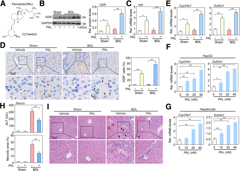
Paricalcitol (VDR agonist, 200 ng/kg) was injected intraperitoneally into bile duct-ligated mice every other day for 5 days. Primary hepatocytes and HepG2 hepatoma cells were transfected with Vdr short hairpin RNA, control short hairpin RNA, Vdr plasmid, control vector, Atg5 small interfering RNA (siRNA), and control siRNA. Liver histology, cell proliferation, and autophagy were evaluated.
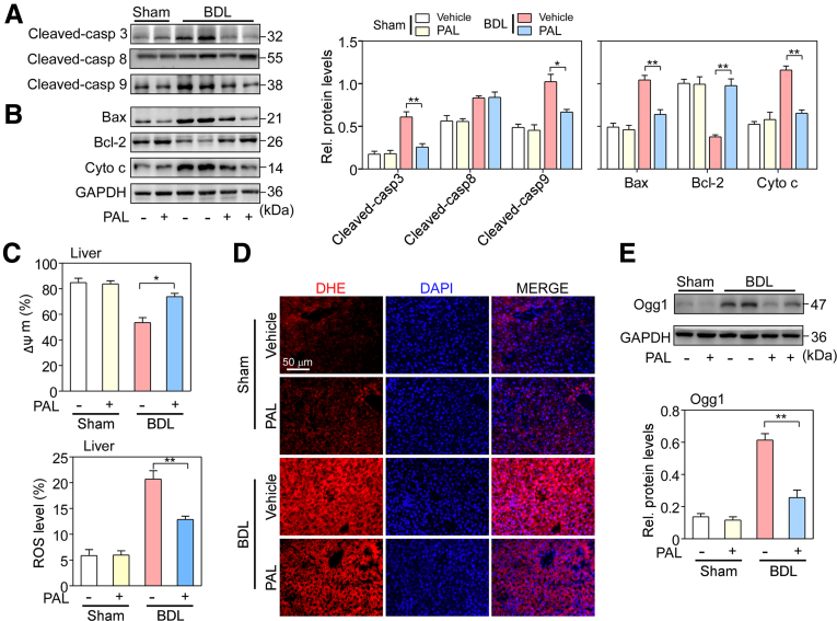
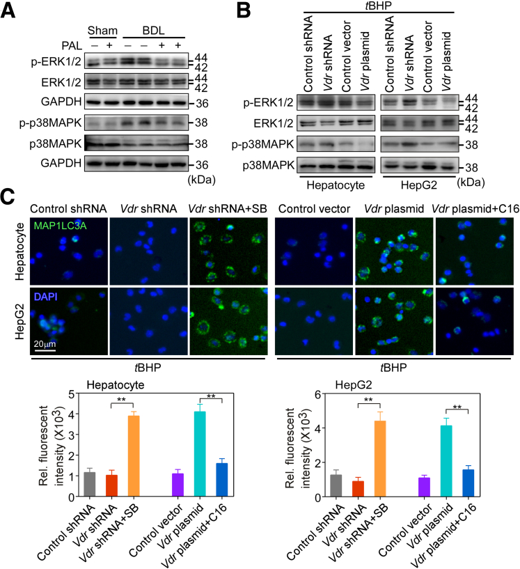
Treatment with the VDR agonist paricalcitol improved liver injury in bile duct-ligated mice by up-regulating VDR expression in hepatocytes, which in turn reduced hepatocyte apoptosis by inhibiting reactive oxygen species (ROS) generation via suppressing the Ras-related C3 botulinum toxin substrate 1/reduced nicotinamide adenine dinucleotide phosphate oxidase 1 pathway. Mechanistically, upon exposure to an ROS-inducing compound, Vdr siRNA contributed to apoptosis, whereas the Vdr overexpression caused resistance to apoptosis. Interestingly, up-regulated VDR expression also increased the generation of autophagosomes and macroautophagic/autophagic flux, which was the underlying mechanism for reduced apoptosis following VDR activation. Autophagy depletion impaired the positive effects of VDR overexpression, whereas autophagy induction was synergystic with VDR overexpression. Importantly, up-regulation of VDR promoted autophagy activation by suppressing the activation of the extracellular signal-regulated kinase (ERK)/p38 mitogen-activated protein kinase (p38MAPK) pathway. Thus, a p38MAPK inhibitor abrogated the Vdr siRNA-induced decrease in autophagy and the Vdr siRNA-induced increase in apoptosis. In contrast, a Mitogen-activated protein kinase kinase (MEK)/ERK activator prevented the enhancement of autophagy and decreased apoptosis following Vdr overexpression. Moreover, the ROS inhibitor N-acetylcystein (NAC) blocked Vdr siRNA-enhanced activation of the ERK/p38MAPK pathway.
VDR activation mitigated liver cholestatic injury by reducing autophagy-dependent hepatocyte apoptosis and suppressing the activation of the ROS-dependent ERK/p38MAPK pathway. Thus, VDR activation may be a potential target for the treatment of cholestatic liver disease.
观察性流行病学研究表明,维生素 D 缺乏与胆汁淤积有关。我们之前曾报道,在胆管细胞中激活维生素 D/维生素 D 受体(VDR)轴通过重塑受损的胆管来减轻胆汁淤积性肝损伤。然而,VDR 在胆汁淤积时在肝细胞中的功能尚不清楚。
每隔一天向胆管结扎小鼠腹膜内注射帕立骨化醇(VDR 激动剂,200ng/kg),共 5 天。用 Vdr 短发夹 RNA、对照短发夹 RNA、Vdr 质粒、对照载体、Atg5 小干扰 RNA(siRNA)和对照 siRNA 转染原代肝细胞和 HepG2 肝癌细胞。评估肝组织学、细胞增殖和自噬。
VDR 激动剂帕立骨化醇通过上调肝细胞中 VDR 的表达来改善胆管结扎小鼠的肝损伤,进而通过抑制 Ras 相关 C3 肉毒杆菌毒素底物 1/还原型烟酰胺腺嘌呤二核苷酸磷酸氧化酶 1 途径抑制活性氧(ROS)生成来减少肝细胞凋亡。在机制上,暴露于诱导 ROS 的化合物后,Vdr siRNA 有助于细胞凋亡,而 Vdr 过表达则通过抑制细胞外信号调节激酶(ERK)/p38 丝裂原激活蛋白激酶(p38MAPK)途径的激活导致对细胞凋亡的抵抗力。有趣的是,上调的 VDR 表达也增加了自噬小体和巨自噬/自噬流的产生,这是 VDR 激活后减少细胞凋亡的潜在机制。自噬耗竭会损害 VDR 过表达的积极作用,而自噬诱导与 VDR 过表达协同作用。重要的是,上调 VDR 通过抑制细胞外信号调节激酶(ERK)/p38 丝裂原激活蛋白激酶(p38MAPK)途径的激活来促进自噬的激活。因此,p38MAPK 抑制剂可阻断 Vdr siRNA 诱导的自噬减少和 Vdr siRNA 诱导的细胞凋亡增加。相反,丝裂原激活蛋白激酶激酶(MEK)/ERK 激活剂可防止 Vdr 过表达后自噬增强和细胞凋亡减少。此外,ROS 抑制剂 N-乙酰半胱氨酸(NAC)阻断 Vdr siRNA 增强的 ERK/p38MAPK 途径的激活。
VDR 激活通过减少自噬依赖性肝细胞凋亡和抑制 ROS 依赖性 ERK/p38MAPK 途径的激活来减轻胆汁淤积性肝损伤。因此,VDR 激活可能是治疗胆汁淤积性肝病的潜在靶点。