Microchemistry, Proteomics, and Lipidomics, Genentech, Inc South San Francisco, CA, USA.
Pharmaceutical Development, Genentech, Inc South San Francisco, CA, USA.
MAbs. 2022 Jan-Dec;14(1):2135183. doi: 10.1080/19420862.2022.2135183.
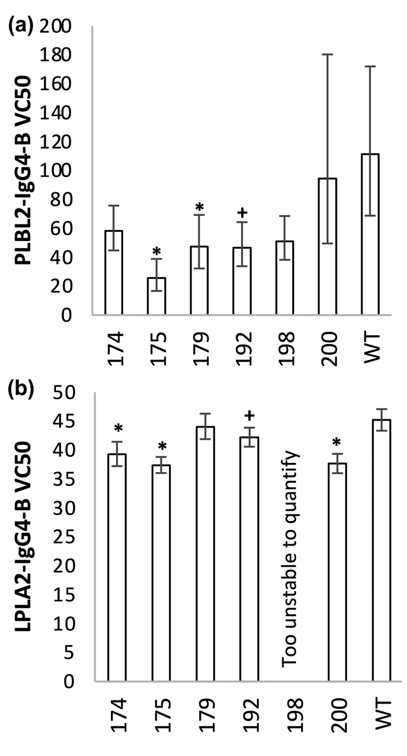
Detection of host cell protein (HCP) impurities is critical to ensuring that recombinant drug products, including monoclonal antibodies (mAbs), are safe. Mechanistic characterization as to how HCPs persist in drug products is important to refining downstream processing. It has been hypothesized that weak lipase-mAb interactions enable HCP lipases to evade drug purification processes. Here, we apply state-of-the-art methods to establish lipase-mAb binding mechanisms. First, the mass spectrometry (MS) approach of fast photochemical oxidation of proteins was used to elucidate putative binding regions. The CH1 domain was identified as a conserved interaction site for IgG1 and IgG4 mAbs against the HCPs phospholipase B-like protein (PLBL2) and lysosomal phospholipase A2 (LPLA2). Rationally designed mutations in the CH1 domain of the IgG4 mAb caused a 3- to 70-fold K reduction against PLBL2 by surface plasmon resonance (SPR). LPLA2-IgG4 mutant complexes, undetected by SPR and studied using native MS collisional dissociation experiments, also showed significant complex disruption, from 16% to 100%. Native MS and ion mobility (IM) determined complex stoichiometries for four lipase-IgG4 complexes and directly interrogated the enrichment of specific lipase glycoforms. Confirmed with time-course and exoglycosidase experiments, deglycosylated lipases prevented binding, and low-molecular-weight glycoforms promoted binding, to mAbs. This work demonstrates the value of integrated biophysical approaches to characterize micromolar affinity complexes. It is the first in-depth structural report of lipase-mAb binding, finding roles for the CH1 domain and lipase glycosylation in mediating binding. The structural insights gained offer new approaches for the bioengineering of cells or mAbs to reduce HCP impurity levels. CAN, Acetonitrile; AMAC, Ammonium acetate; BFGS, Broyden-Fletcher-Goldfarb-Shanno; CHO, Chinese Hamster Ovary; K, Dissociation constant; DTT, Dithiothreitol; ELISA, Enzyme-linked immunosorbent assay; FPOP, Fast photochemical oxidation of proteins; FA, Formic acid; F(ab'), Fragment antibodies; HCP, Host cell protein; IgG, Immunoglobulin; IM, Ion mobility; LOD, Lower limit of detection; LPLA2, Lysosomal phospholipase A2; Man, Mannose; MS, Mass spectrometry; MeOH, Methanol; MST, Microscale thermophoresis; mAbs, Monoclonal antibodies; PPT1, Palmitoyl protein thioesterase; ppm, Parts per million; PLBL2, Phospholipase B-like protein; PLD3, Phospholipase D3; PS-20, Polysorbate-20; SP, Sphingomyelin phosphodiesterase; SPR, Surface plasmon resonance; TFA, Trifluoroacetic acid.
宿主细胞蛋白(HCP)杂质的检测对于确保重组药物产品(包括单克隆抗体(mAbs))的安全性至关重要。了解 HCP 如何在药物产品中持续存在的机制特征对于改进下游加工过程非常重要。据推测,弱的脂肪酶-mAb 相互作用使 HCP 脂肪酶能够逃避药物纯化过程。在这里,我们应用最先进的方法来建立脂肪酶-mAb 结合机制。首先,使用快速光化学氧化蛋白质的质谱(MS)方法来阐明假定的结合区域。CH1 结构域被确定为针对 HCP 磷脂酶 B 样蛋白(PLBL2)和溶酶体磷脂酶 A2(LPLA2)的 IgG1 和 IgG4 mAb 的保守相互作用位点。通过表面等离子体共振(SPR),对 IgG4 mAb 的 CH1 结构域进行合理设计的突变导致对 PLBL2 的 K 值降低了 3 到 70 倍。SPR 无法检测到的 LPLA2-IgG4 突变体复合物,并使用天然 MS 碰撞解离实验进行了研究,也显示出明显的复合物破坏,从 16%到 100%。天然 MS 和离子迁移(IM)确定了四个脂肪酶-IgG4 复合物的复合物化学计量,并直接检测了特定脂肪酶糖型的富集。通过时间过程和外糖苷酶实验证实,去糖基化的脂肪酶阻止结合,而低分子量的糖型促进与 mAb 的结合。这项工作证明了综合生物物理方法用于表征微摩尔亲和力复合物的价值。这是首次深入研究脂肪酶-mAb 结合的结构报告,发现 CH1 结构域和脂肪酶糖基化在介导结合中的作用。获得的结构见解为降低 HCP 杂质水平的细胞或 mAb 的生物工程提供了新的方法。CAN,乙腈;AMAC,乙酸铵;BFGS,Broyden-Fletcher-Goldfarb-Shanno;CHO,中国仓鼠卵巢;K,离解常数;DTT,二硫苏糖醇;ELISA,酶联免疫吸附测定;FPOP,快速光化学氧化蛋白质;FA,甲酸;F(ab'),片段抗体;HCP,宿主细胞蛋白;IgG,免疫球蛋白;IM,离子迁移;LOD,检测下限;LPLA2,溶酶体磷脂酶 A2;甘露糖;MS,质谱;MeOH,甲醇;MST,微尺度热泳;mAbs,单克隆抗体;PPT1,棕榈酰蛋白硫酯酶;ppm,百万分之几;PLBL2,磷脂酶 B 样蛋白;PLD3,磷脂酶 D3;PS-20,聚山梨酯 20;SP,鞘磷脂磷酸二酯酶;SPR,表面等离子体共振;TFA,三氟乙酸。