Chung Jin-Yong, Park Ji-Eun, Kim Yoon-Jae, Lee Seung-Jin, Yu Wook-Joon, Kim Jong-Min
Department of Anatomy and Cell Biology, College of Medicine, Dong-A University, Busan 49201, Korea.
Developmental and Reproductive Toxicology Research Group, Korea Institute of Toxicology, Daejeon 34114, Korea.
Dev Reprod. 2022 Sep;26(3):99-105. doi: 10.12717/DR.2022.26.3.99. Epub 2022 Sep 30.
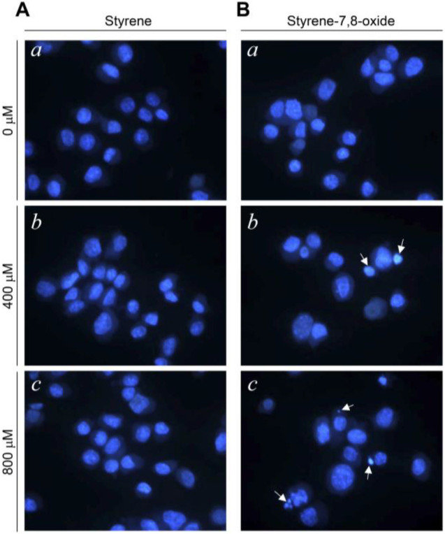
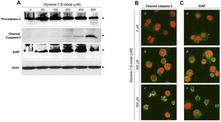
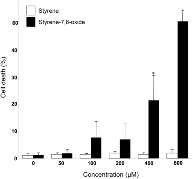
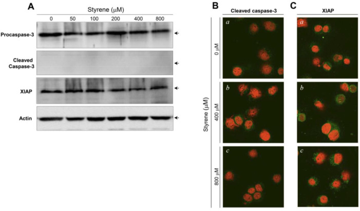
Styrene is the precursor of polystyrene. Human exposure to styrene could occur in occupational and residential settings and via food intake. Styrene is metabolized to styrene-7,8-oxide by cytochrome P450 enzyme. In the present study, we investigated the cytotoxicity mediated by styrene and styrene-7,8-oxide in TM3 testicular Leydig cells . We first monitored the nuclear fragmentation in Leydig cells after exposure to styrene or styrene-7,8-oxide. Hoechst 33258 cell staining showed that styrene exposure in TM3 Leydig cells did not exhibit nuclear fragmentation at any concentration. In contrast, nuclear fragmentation was seen in styrene-7,8-oxide-exposed cells. These results indicate that cytotoxicity-mediated cell death in Leydig cells is more susceptible to styrene-7,8-oxide than to styrene. Following styrene treatment, procaspase-3 and XIAP protein levels did not show significant changes, and cleaved (active) forms of caspase-3 were not detected. Consistent with the western blot results, the active forms of caspase-3 and XIAP proteins were not prominently altered in the cytoplasm of cells treated with styrene. In contrast to styrene, styrene-7,8-oxide induced cell death in an apoptotic fashion, as seen in caspase-3 activation and increased the expression of XIAP proteins. Taken together, the results obtained in this study demonstrate a fundamental idea that Leydig cells are capable of protecting themselves from cytotoxicity-mediated apoptosis as a result of styrene exposure . It remains unclear whether the steroid-producing function, i.e., steroidogenesis, of Leydig cells is also unaffected by exposure to styrene. Therefore, further studies are needed to elucidate the endocrine disrupting potential of styrene in Leydig cells.
苯乙烯是聚苯乙烯的前体。人类可通过职业和居住环境以及食物摄入接触苯乙烯。苯乙烯通过细胞色素P450酶代谢为苯乙烯-7,8-氧化物。在本研究中,我们调查了苯乙烯和苯乙烯-7,8-氧化物在TM3睾丸间质细胞中介导的细胞毒性。我们首先监测了间质细胞在接触苯乙烯或苯乙烯-7,8-氧化物后的核碎裂情况。Hoechst 33258细胞染色显示,TM3间质细胞暴露于苯乙烯后,在任何浓度下均未出现核碎裂。相比之下,在暴露于苯乙烯-7,8-氧化物的细胞中可见核碎裂。这些结果表明,间质细胞中由细胞毒性介导的细胞死亡对苯乙烯-7,8-氧化物比对苯乙烯更敏感。苯乙烯处理后,原半胱天冬酶-3和XIAP蛋白水平未显示出显著变化,且未检测到裂解(活性)形式的半胱天冬酶-3。与蛋白质印迹结果一致,在用苯乙烯处理的细胞的细胞质中,半胱天冬酶-3和XIAP蛋白的活性形式没有明显改变。与苯乙烯不同,苯乙烯-7,8-氧化物以凋亡方式诱导细胞死亡,如半胱天冬酶-3激活所示,并增加了XIAP蛋白的表达。综上所述,本研究获得的结果证明了一个基本观点,即间质细胞能够保护自身免受苯乙烯暴露导致的细胞毒性介导的凋亡。目前尚不清楚间质细胞的类固醇生成功能,即类固醇合成,是否也不受苯乙烯暴露的影响。因此,需要进一步研究以阐明苯乙烯在间质细胞中的内分泌干扰潜力。