Schmidt Ryder M, Hara Daiki, Vega Jorge D, Abuhaija Marwan B, Tao Wensi, Dogan Nesrin, Pollack Alan, Ford John C, Shi Junwei
Department of Radiation Oncology, Miller School of Medicine, University of Miami, Miami, FL 33136, USA.
Department of Biomedical Engineering, University of Miami, Coral Cables, FL 33146, USA.
Pharmaceutics. 2022 Oct 17;14(10):2205. doi: 10.3390/pharmaceutics14102205.
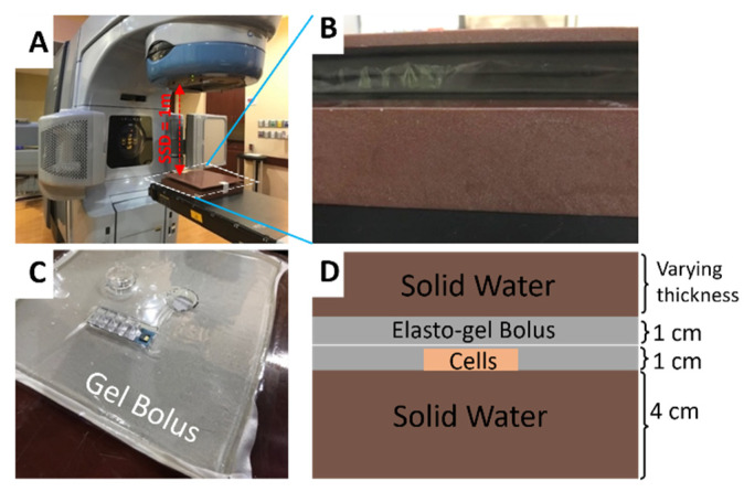
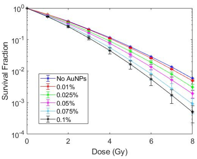
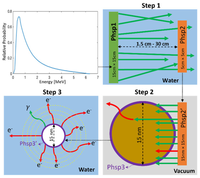
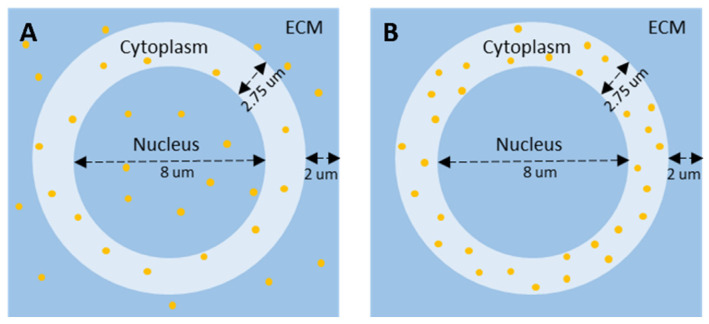
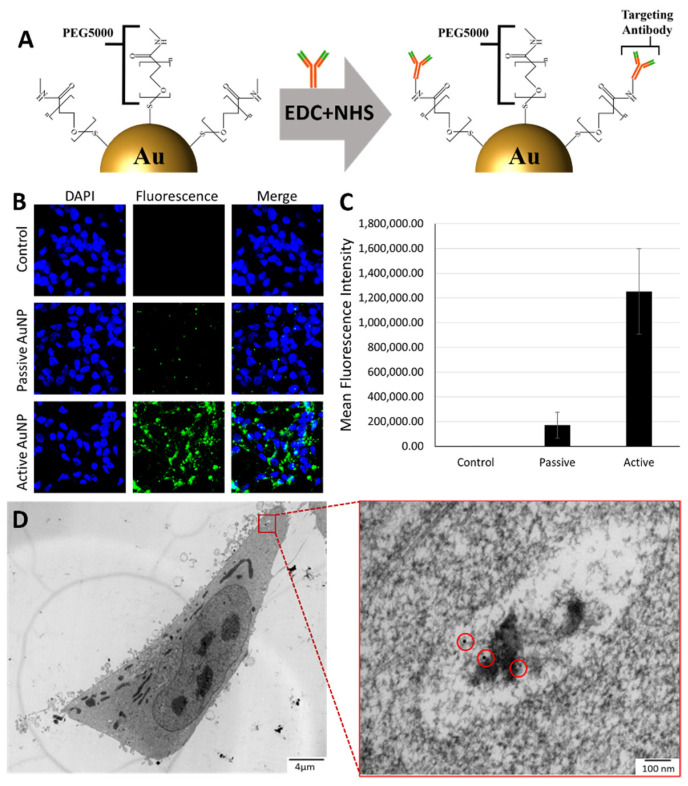
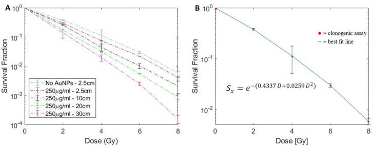
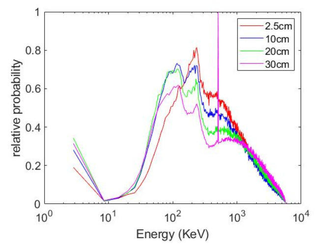
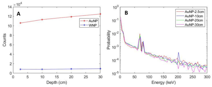
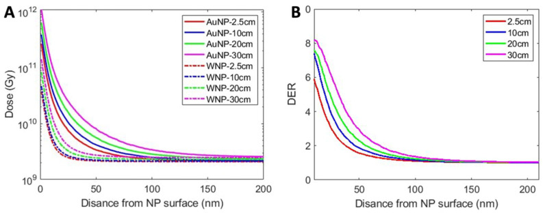
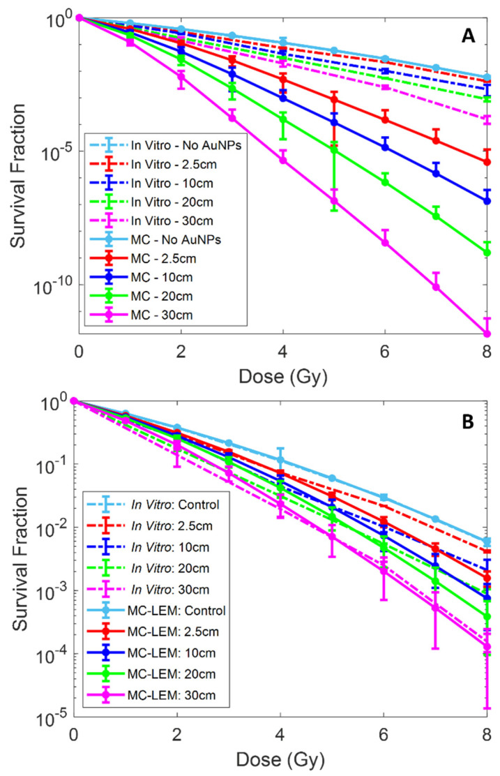
Active targeting gold nanoparticles (AuNPs) are a very promising avenue for cancer treatment with many publications on AuNP mediated radiosensitization at kilovoltage (kV) photon energies. However, uncertainty on the effectiveness of AuNPs under clinically relevant megavoltage (MV) radiation energies hinders the clinical translation of AuNP-assisted radiation therapy (RT) paradigm. The aim of this study was to investigate radiosensitization mediated by PSMA-targeted AuNPs irradiated by a 6 MV radiation beam at different depths to explore feasibility of AuNP-assisted prostate cancer RT under clinically relevant conditions. PSMA-targeted AuNPs (PSMA-AuNPs) were synthesized by conjugating PSMA antibodies onto PEGylated AuNPs through EDC/NHS chemistry. Confocal fluorescence microscopy was used to verify the active targeting of the developed PSMA-AuNPs. Transmission electron microscopy (TEM) was used to demonstrate the intracellular biodistribution of PSMA-AuNPs. LNCaP prostate cancer cells treated with PSMA-AuNPs were irradiated on a Varian 6 MV LINAC under varying depths (2.5 cm, 10 cm, 20 cm, 30 cm) of solid water. Clonogenic assays were carried out to determine the in vitro cell survival fractions. A Monte Carlo (MC) model developed on TOPAS platform was then employed to determine the nano-scale radial dose distribution around AuNPs, which was subsequently used to predict the radiation dose response of LNCaP cells treated with AuNPs. Two different cell models, with AuNPs located within the whole cell or only in the cytoplasm, were used to assess how the intracellular PSMA-AuNP biodistribution impacts the prostate cancer radiosensitization. Then, MC-based microdosimetry was combined with the local effect model (LEM) to calculate cell survival fraction, which was benchmarked against the in vitro clonogenic assays at different depths. In vitro clonogenic assay of LNCaP cells demonstrated the depth dependence of AuNP radiosensitization under clinical megavoltage beams, with sensitization enhancement ratio (SER) of 1.14 ± 0.03 and 1.55 ± 0.05 at 2.5 cm depth and 30 cm depth, respectively. The MC microdosimetry model showed the elevated percent of low-energy photons in the MV beams at greater depth, consequently resulting in increased dose enhancement ratio (DER) of AuNPs with depth. The AuNP-induced DER reached ~5.7 and ~8.1 at depths of 2.5 cm and 30 cm, respectively. Microdosimetry based LEM accurately predicted the cell survival under 6 MV beams at different depths, for the cell model with AuNPs placed only in the cell cytoplasm. TEM results demonstrated the distribution of PSMA-AuNPs in the cytoplasm, confirming the accuracy of MC microdosimetry based LEM with modelled AuNPs distributed within the cytoplasm. We conclude that AuNP radiosensitization can be achieved under megavoltage clinical radiotherapy energies with a dependence on tumor depth. Furthermore, the combination of Monte Carlo microdosimetry and LEM will be a valuable tool to assist with developing AuNP-aided radiotherapy paradigm and drive clinical translation.
主动靶向金纳米颗粒(AuNPs)是癌症治疗中一条非常有前景的途径,有许多关于千伏(kV)光子能量下AuNP介导的放射增敏作用的出版物。然而,在临床相关的兆伏(MV)辐射能量下AuNPs有效性的不确定性阻碍了AuNP辅助放射治疗(RT)模式的临床转化。本研究的目的是研究在不同深度用6 MV辐射束照射PSMA靶向的AuNPs介导的放射增敏作用,以探索在临床相关条件下AuNP辅助前列腺癌RT的可行性。通过EDC/NHS化学方法将PSMA抗体偶联到聚乙二醇化的AuNPs上,合成了PSMA靶向的AuNPs(PSMA-AuNPs)。共聚焦荧光显微镜用于验证所制备的PSMA-AuNPs的主动靶向性。透射电子显微镜(TEM)用于证明PSMA-AuNPs在细胞内的生物分布。用PSMA-AuNPs处理的LNCaP前列腺癌细胞在瓦里安6 MV直线加速器上,在不同深度(2.5 cm、10 cm、20 cm、30 cm)的固体水模体下进行照射。进行克隆形成试验以确定体外细胞存活分数。然后采用在TOPAS平台上开发的蒙特卡罗(MC)模型来确定AuNPs周围的纳米级径向剂量分布,随后用于预测用AuNPs处理的LNCaP细胞的辐射剂量反应。使用两种不同的细胞模型(AuNPs位于整个细胞内或仅位于细胞质中)来评估细胞内PSMA-AuNP生物分布如何影响前列腺癌放射增敏作用。然后,基于MC的微剂量学与局部效应模型(LEM)相结合来计算细胞存活分数,并与不同深度的体外克隆形成试验结果进行对比。LNCaP细胞的体外克隆形成试验证明了在临床兆伏束下AuNP放射增敏作用的深度依赖性,在2.5 cm深度和30 cm深度时,增敏增强比(SER)分别为1.14±0.03和1.55±0.05。MC微剂量学模型显示,在更深的深度,MV束中低能光子的百分比升高,因此导致AuNPs的剂量增强比(DER)随深度增加。在2.5 cm和30 cm深度时,AuNP诱导的DER分别达到约5.7和约8.1。基于微剂量学的LEM准确预测了在不同深度的6 MV束下,AuNPs仅位于细胞质中的细胞模型的细胞存活情况。TEM结果证明了PSMA-AuNPs在细胞质中的分布,证实了基于MC微剂量学的LEM(其中AuNPs分布在细胞质中)的准确性。我们得出结论,在兆伏临床放射治疗能量下可以实现AuNP放射增敏作用,且依赖于肿瘤深度。此外,蒙特卡罗微剂量学和LEM的结合将是协助开发AuNP辅助放射治疗模式并推动临床转化的有价值工具。