Department of Biotechnology, Technische Universität Braunschweig, Spielmannstraße 7, 38106 Braunschweig, Germany.
Recombinant Protein Expression Platform, Helmholtz Centre for Infection Research, Inhoffenstraße 7, 38124 Braunschweig, Germany.
Viruses. 2022 Sep 20;14(10):2087. doi: 10.3390/v14102087.
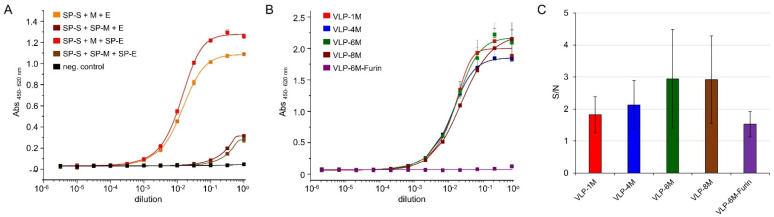
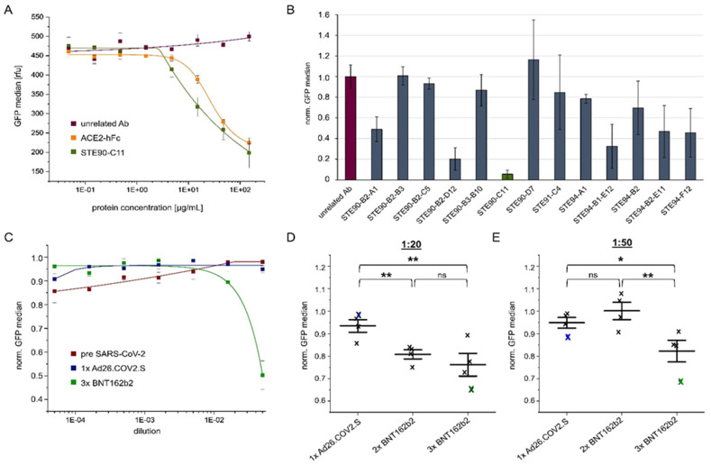
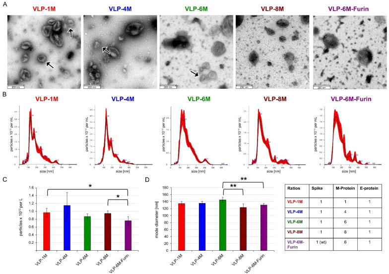
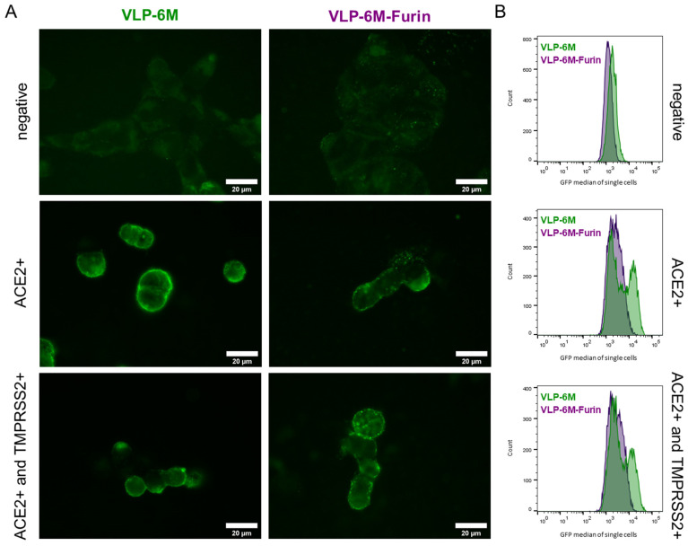
Virus-like particles (VLPs) resemble authentic virus while not containing any genomic information. Here, we present a fast and powerful method for the production of SARS-CoV-2 VLP in insect cells and the application of these VLPs to evaluate the inhibition capacity of monoclonal antibodies and sera of vaccinated donors. Our method avoids the baculovirus-based approaches commonly used in insect cells by employing direct plasmid transfection to co-express SARS-CoV-2 envelope, membrane, and spike protein that self-assemble into VLPs. After optimization of the expression plasmids and vector ratios, VLPs with an ~145 nm diameter and the typical "Corona" aura were obtained, as confirmed by nanoparticle tracking analysis (NTA) and transmission electron microscopy (TEM). Fusion of the membrane protein to GFP allowed direct quantification of binding inhibition to angiotensin II-converting enzyme 2 (ACE2) on cells by therapeutic antibody candidates or sera from vaccinated individuals. Neither VLP purification nor fluorescent labeling by secondary antibodies are required to perform these flow cytometric assays.
病毒样颗粒(VLPs)类似于真实的病毒,但不包含任何基因组信息。在这里,我们提出了一种在昆虫细胞中生产 SARS-CoV-2 VLP 的快速而强大的方法,并应用这些 VLP 来评估单克隆抗体和疫苗接种供体血清的抑制能力。我们的方法避免了昆虫细胞中常用的基于杆状病毒的方法,而是通过直接质粒转染共表达 SARS-CoV-2 包膜、膜和刺突蛋白,这些蛋白自行组装成 VLPs。通过对表达质粒和载体比例的优化,获得了直径约 145nm 且具有典型“日冕”光环的 VLPs,这通过纳米颗粒跟踪分析(NTA)和透射电子显微镜(TEM)得到了证实。膜蛋白与 GFP 的融合允许通过候选治疗性抗体或疫苗接种个体的血清直接定量测定与细胞上的血管紧张素转化酶 2(ACE2)的结合抑制。进行这些流式细胞术测定不需要 VLP 纯化或二级抗体的荧光标记。