Department of Orthopedics, Binhai County People's Hospital, Jiangsu Province, China.
Department of Geriatrics, Binhai County People's Hospital, Binhai, Jiangsu Province, China.
Immun Inflamm Dis. 2022 Nov;10(11):e714. doi: 10.1002/iid3.714.
Macrophages are the only inflammatory cells that can penetrate the closed nucleus pulposus and their polarization plays an important role in intervertebral disc degeneration (IVDD). This paper attempted to investigate the pathogenesis of IVDD by altering the polarization state of macrophages.
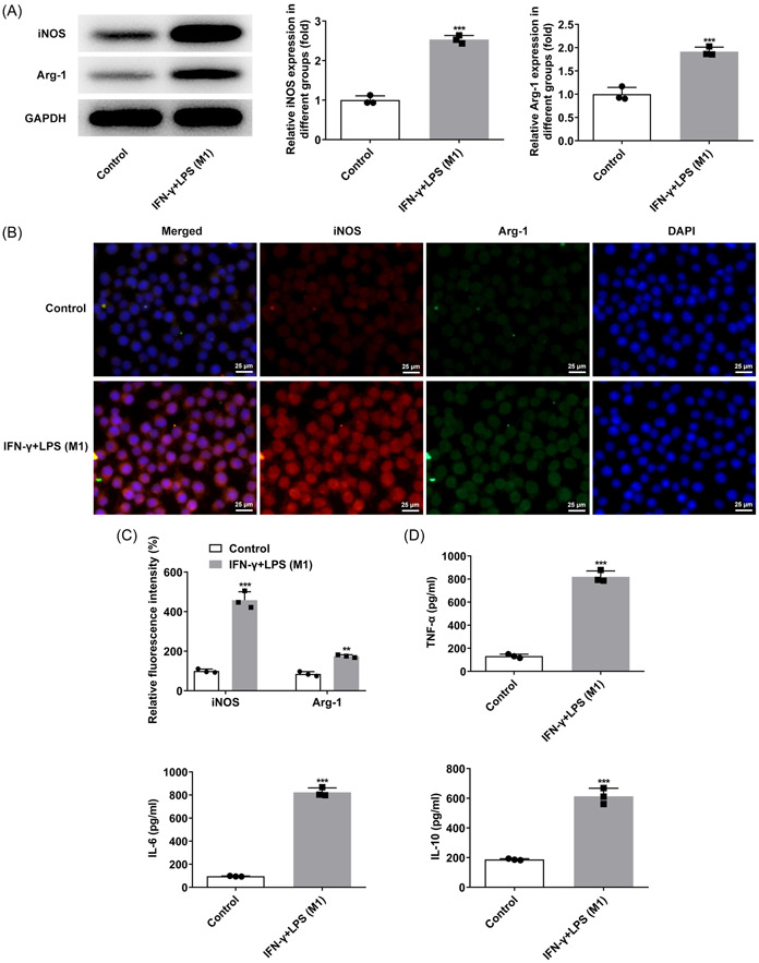
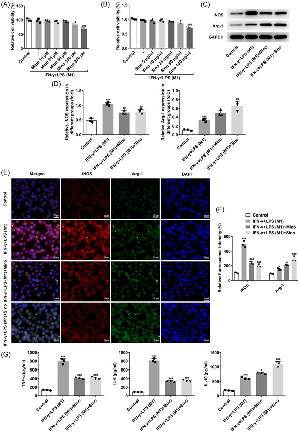
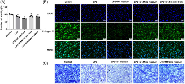
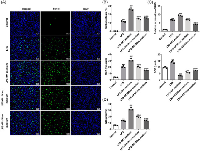
Macrophage RAW264.7 cells were induced by interferonγ (IFN-γ) and lipopolysaccharide (LPS). The polarization of RAW264.7 cells was estimated by western blot and immunofluorescence. The expressions of inflammatory factors were detected by ELISA. Subsequently, RAW264.7 cells were treated with different concentrations of minocycline (Mino) and sinomenine (Sino), followed by the assessment of cell viability with cell counting kit-8 kit. Then, RAW264.7 cell culture medium was collected for the culture of human nucleus pulposus cells (NPCs). Toluidine blue staining and type II collagen staining were applied to assay the level of type II collagen. The cell apoptosis, oxidative stress, and nitric oxide (NO) level were appraised by TUNEL, oxidative stress kits and NO kit, respectively. Western blot was employed to test the levels of apoptosis- and oxidative stress-related proteins.
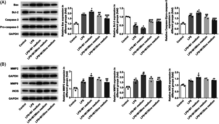
IFN-γ and LPS could induce M1 polarization of RAW264.7 cells. Mino and Sino could reduce the polarization of RAW264.7 cells toward M1. M1-polarized medium inhibited LPS-induced activity, inflammation, and damage of NPCs, which were enhanced by Mino and Sino in medium.
M1 polarization of macrophages promoted LPS-induced inflammation and damage of NPCs.
巨噬细胞是唯一能够穿透封闭的髓核的炎症细胞,其极化在椎间盘退变(IVDD)中起着重要作用。本文试图通过改变巨噬细胞的极化状态来研究 IVDD 的发病机制。
用干扰素γ(IFN-γ)和脂多糖(LPS)诱导巨噬细胞 RAW264.7 细胞。通过 Western blot 和免疫荧光法评估 RAW264.7 细胞的极化状态。通过 ELISA 检测炎症因子的表达。随后,用不同浓度的米诺环素(Mino)和青藤碱(Sino)处理 RAW264.7 细胞,用细胞计数试剂盒-8 检测细胞活力。然后收集 RAW264.7 细胞培养上清液培养人髓核细胞(NPCs)。甲苯胺蓝染色和 II 型胶原染色检测 II 型胶原水平。通过 TUNEL 检测细胞凋亡,氧化应激试剂盒和 NO 试剂盒检测氧化应激和一氧化氮(NO)水平。Western blot 检测凋亡和氧化应激相关蛋白的水平。
IFN-γ 和 LPS 可诱导 RAW264.7 细胞向 M1 极化。米诺环素和青藤碱可减少 RAW264.7 细胞向 M1 极化。M1 极化的培养基抑制了 LPS 诱导的 NPCs 的活性、炎症和损伤,而米诺环素和青藤碱在培养基中增强了这些作用。
巨噬细胞的 M1 极化促进了 LPS 诱导的 NPCs 的炎症和损伤。