Department of Biomedical Engineering, Boston University, Boston, Massachusetts, United States of America.
Department of Numerical Analysis and Scientific Computing, Simula Research Laboratory, Oslo, Norway.
PLoS Biol. 2022 Oct 27;20(10):e3001440. doi: 10.1371/journal.pbio.3001440. eCollection 2022 Oct.
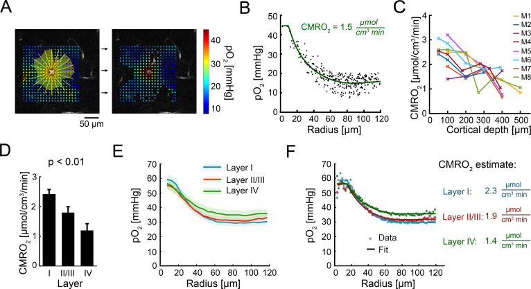
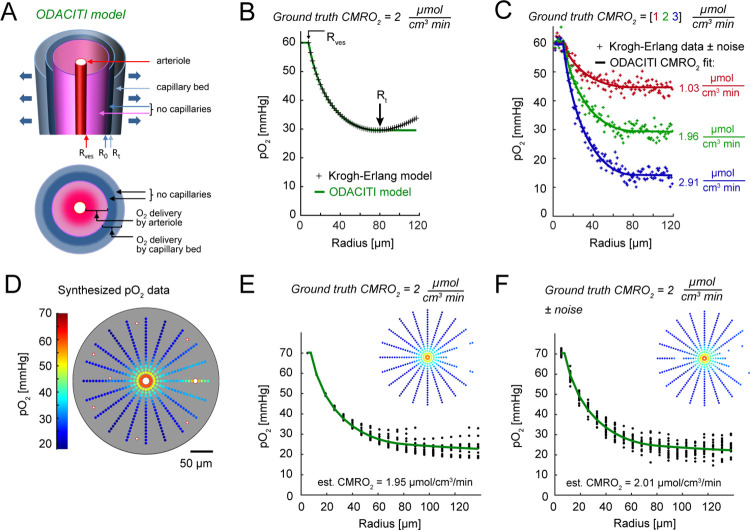
The cerebral cortex is organized in cortical layers that differ in their cellular density, composition, and wiring. Cortical laminar architecture is also readily revealed by staining for cytochrome oxidase-the last enzyme in the respiratory electron transport chain located in the inner mitochondrial membrane. It has been hypothesized that a high-density band of cytochrome oxidase in cortical layer IV reflects higher oxygen consumption under baseline (unstimulated) conditions. Here, we tested the above hypothesis using direct measurements of the partial pressure of O2 (pO2) in cortical tissue by means of 2-photon phosphorescence lifetime microscopy (2PLM). We revisited our previously developed method for extraction of the cerebral metabolic rate of O2 (CMRO2) based on 2-photon pO2 measurements around diving arterioles and applied this method to estimate baseline CMRO2 in awake mice across cortical layers. To our surprise, our results revealed a decrease in baseline CMRO2 from layer I to layer IV. This decrease of CMRO2 with cortical depth was paralleled by an increase in tissue oxygenation. Higher baseline oxygenation and cytochrome density in layer IV may serve as an O2 reserve during surges of neuronal activity or certain metabolically active brain states rather than reflecting baseline energy needs. Our study provides to our knowledge the first quantification of microscopically resolved CMRO2 across cortical layers as a step towards better understanding of brain energy metabolism.
大脑皮层组织在细胞密度、成分和连接上存在差异,其皮层层结构也可以通过对细胞色素氧化酶(细胞呼吸电子传递链中的最后一种酶,位于线粒体内膜)的染色轻易地显现出来。有人假设,皮层 IV 层中细胞色素氧化酶的高密度带反映了在基线(未受刺激)条件下更高的耗氧量。在这里,我们使用 2 光子磷光寿命显微镜(2PLM)直接测量皮质组织中的部分氧分压(pO2),对上述假设进行了测试。我们重新使用了之前开发的方法,基于在潜水小动脉周围的 2 光子 pO2 测量来提取大脑耗氧量(CMRO2),并将该方法应用于估计清醒小鼠在整个皮层层的基线 CMRO2。令我们惊讶的是,我们的结果显示从皮层 I 层到皮层 IV 层,基线 CMRO2 下降。这种 CMRO2 随皮层深度的降低与组织氧合作用的增加相平行。IV 层中更高的基线氧合作用和细胞色素密度可能是神经元活动激增或某些代谢活跃的脑状态下的 O2 储备,而不是反映基线能量需求。我们的研究提供了我们所知的首次对皮层各层之间微观分辨率的 CMRO2 的定量,这是更好地理解大脑能量代谢的一个步骤。