Escandón Joseph M, Mohammad Arbab, Matsui Chihiro, Tanaka Takakuni, Wei-Kai Lao William, Yoshitsugu Hattori, Matsui Yuki, Mizuno Hiroshi
Division of Plastic and Reconstructive Surgery, Strong Memorial Hospital, University of Rochester Medical Center, Rochester, N.Y.
Aarupadai Veedu Medical College and Hospital, Puducherry, India.
Plast Reconstr Surg Glob Open. 2022 Nov 1;10(11):e4583. doi: 10.1097/GOX.0000000000004583. eCollection 2022 Nov.
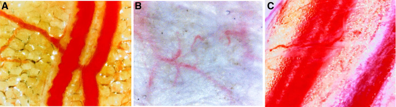
Indocyanine green, ultrasonography, and handheld Doppler can be used to evaluate blood flow at the donor and recipient site during microvascular reconstruction. However, these methods do not provide direct visualization and assessment of real-time blood flow. Video-capillaroscopy has been shown to be useful in clinical practice to assess microcirculation in rheumatologic disorders. In this report we used video-capillaroscopy to assess different tissue components involved in microvascular surgery. Seven patients who underwent head and neck oncologic microvascular reconstruction between November 2021 and February 2022 were included in this study. Video-capillaroscopy (GOKO-BscanZD, GOKO Imaging Devices Co., Ltd., Japan) was used to evaluate the donor-site and recipient-site tissue components. Optimal red blood cell movement was graded with a score of four, while no flow was graded with a score of 0. Seven myocutaneous flaps and seven recipient sites were evaluated. For the donor-site, our analysis demonstrated a significantly higher video-capillaroscopy quality for skin (3.43), adipose tissue (3.7) and perforators (3.7) when compared with muscle (0.429), muscle fascia (0.857), and de-epithelialized skin (1) ( < 0.001). For the recipient-site, a significantly higher video-capillaroscopy quality for skin (2.7), adipose tissue (3.5), and the periosteum (2.1) was noted when compared with muscle (0) ( < 0.001). Video-capillaroscopy efficiency is limited in the muscular component and injured (de-epithelialized) skin surface areas of flaps. Herein, we provide evidence that assessment of flap perfusion with video-capillaroscopy can be reliably achieved in the skin, periosteum, perforators, and adipose tissue. Video-capillaroscopy is expected to be applied for intraoperative real-time blood flow evaluation.
吲哚菁绿、超声检查和手持多普勒可用于评估微血管重建过程中供体和受体部位的血流情况。然而,这些方法无法直接可视化和评估实时血流。视频毛细血管镜已被证明在临床实践中对评估风湿性疾病的微循环有用。在本报告中,我们使用视频毛细血管镜评估微血管手术中涉及的不同组织成分。本研究纳入了2021年11月至2022年2月期间接受头颈肿瘤微血管重建的7例患者。使用视频毛细血管镜(GOKO-BscanZD,日本GOKO成像设备有限公司)评估供体部位和受体部位的组织成分。最佳红细胞运动评分为4分,无血流评分为0分。评估了7个肌皮瓣和7个受体部位。对于供体部位,我们的分析表明,与肌肉(0.429)、肌筋膜(0.857)和去上皮化皮肤(1)相比,皮肤(3.43)、脂肪组织(3.7)和穿支血管(3.7)的视频毛细血管镜质量显著更高(<0.001)。对于受体部位,与肌肉(0)相比,皮肤(2.7)、脂肪组织(3.5)和骨膜(2.1)的视频毛细血管镜质量显著更高(<0.001)。视频毛细血管镜在皮瓣的肌肉成分和受伤(去上皮化)皮肤表面区域的效率有限。在此,我们提供证据表明,通过视频毛细血管镜评估皮瓣灌注在皮肤、骨膜、穿支血管和脂肪组织中可以可靠地实现。视频毛细血管镜有望用于术中实时血流评估。