• 文献检索
  • 文档翻译
  • 深度研究
  • 学术资讯
  • Suppr Zotero 插件Zotero 插件
  • 邀请有礼
  • 套餐&价格
  • 历史记录
应用&插件
Suppr Zotero 插件Zotero 插件浏览器插件Mac 客户端Windows 客户端微信小程序
定价
高级版会员购买积分包购买API积分包
服务
文献检索文档翻译深度研究API 文档MCP 服务
关于我们
关于 Suppr公司介绍联系我们用户协议隐私条款
关注我们

Suppr 超能文献

核心技术专利:CN118964589B侵权必究
粤ICP备2023148730 号-1Suppr @ 2026

文献检索

告别复杂PubMed语法,用中文像聊天一样搜索,搜遍4000万医学文献。AI智能推荐,让科研检索更轻松。

立即免费搜索

文件翻译

保留排版,准确专业,支持PDF/Word/PPT等文件格式,支持 12+语言互译。

免费翻译文档

深度研究

AI帮你快速写综述,25分钟生成高质量综述,智能提取关键信息,辅助科研写作。

立即免费体验

眼球内收时组织特性变化对人视神经束缚影响的有限元建模

Finite element modeling of effects of tissue property variation on human optic nerve tethering during adduction.

机构信息

Department of Ophthalmology, Stein Eye Institute, University of California, Los Angeles, Los Angeles, CA, 90095-7002, USA.

Department of Bioengineering, University of California, Los Angeles, Los Angeles, CA, USA.

出版信息

Sci Rep. 2022 Nov 8;12(1):18985. doi: 10.1038/s41598-022-22899-2.

DOI:10.1038/s41598-022-22899-2
PMID:36347907
原文链接:https://pmc.ncbi.nlm.nih.gov/articles/PMC9643519/
Abstract

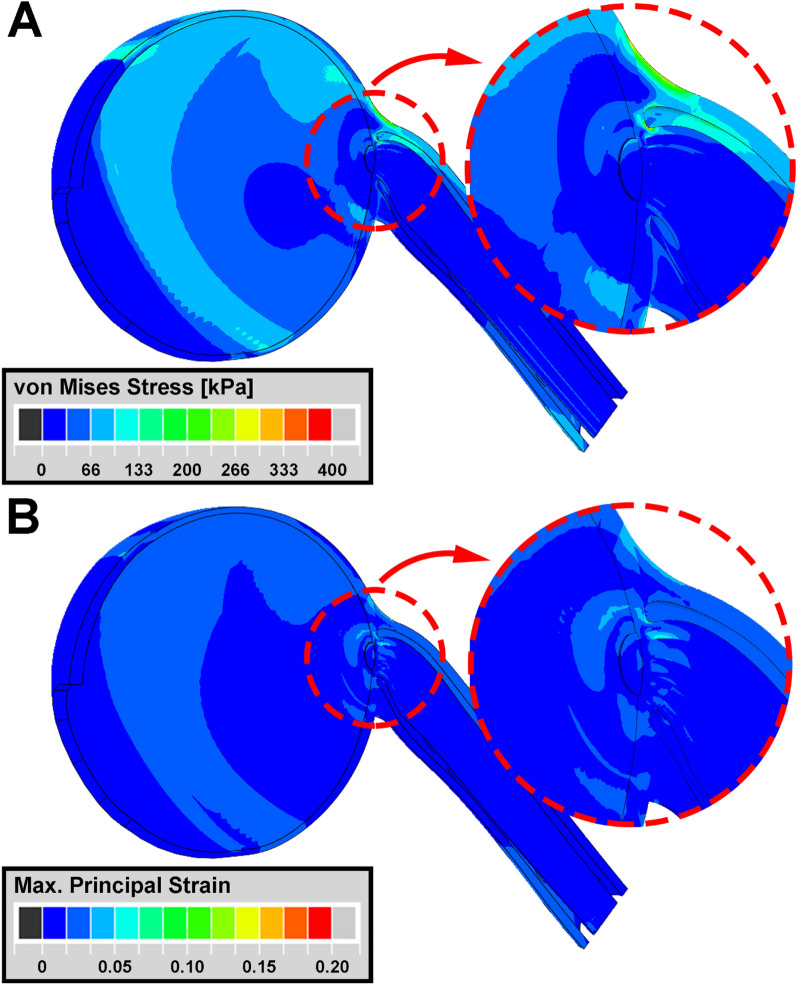
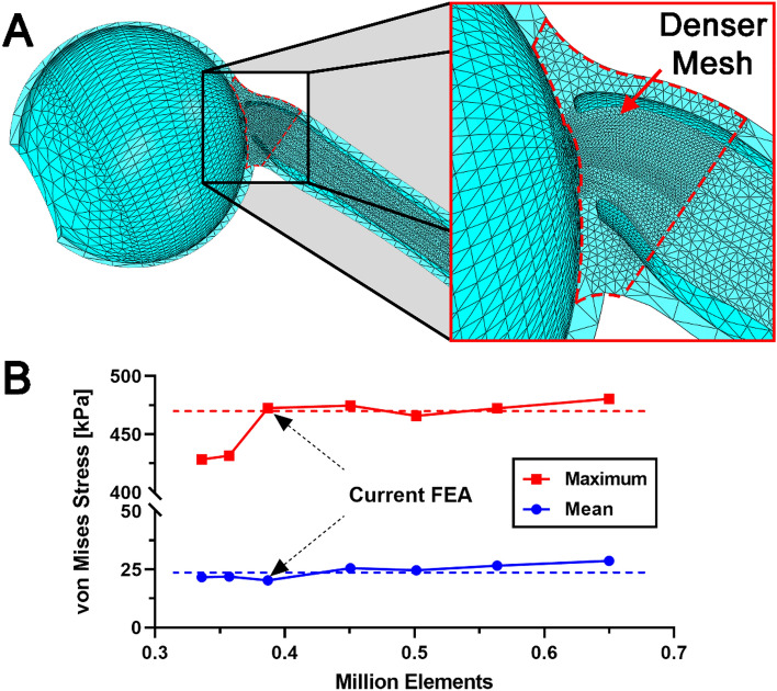
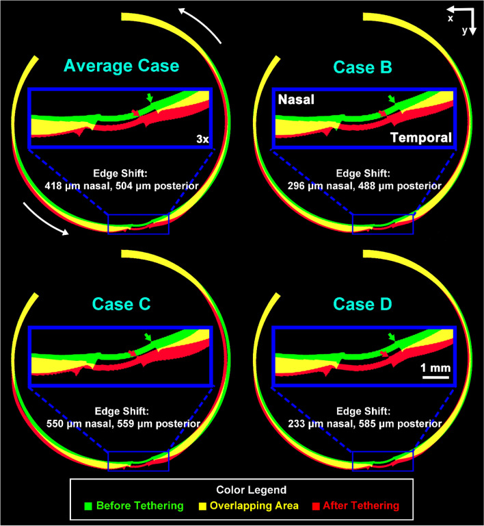
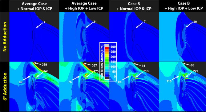
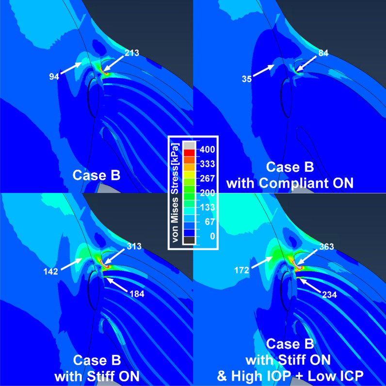
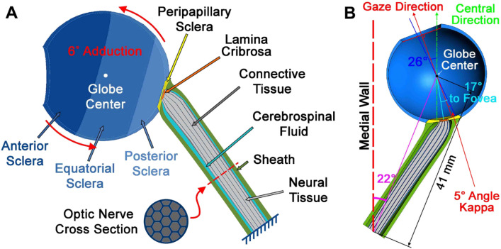
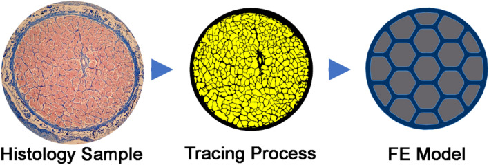
Tractional tethering by the optic nerve (ON) on the eye as it rotates towards the midline in adduction is a significant ocular mechanical load and has been suggested as a cause of ON damage induced by repetitive eye movements. We designed an ocular finite element model (FEM) simulating 6° incremental adduction beyond the initial configuration of 26° adduction that is the observed threshold for ON tethering. This FEM permitted sensitivity analysis of ON tethering using observed material property variations in measured hyperelasticity of the anterior, equatorial, posterior, and peripapillary sclera; and the ON and its sheath. The FEM predicted that adduction beyond the initiation of ON tethering concentrates stress and strain on the temporal side of the optic disc and peripapillary sclera, the ON sheath junction with the sclera, and retrolaminar ON neural tissue. However, some unfavorable combinations of tissue properties within the published ranges imposed higher stresses in these regions. With the least favorable combinations of tissue properties, adduction tethering was predicted to stress the ON junction and peripapillary sclera more than extreme conditions of intraocular and intracranial pressure. These simulations support the concept that ON tethering in adduction could induce mechanical stresses that might contribute to ON damage.

摘要

眼球向中线内收时视神经(ON)被牵引,这是一种显著的眼球机械负荷,被认为是重复眼球运动引起 ON 损伤的原因。我们设计了一个眼球有限元模型(FEM),模拟了 6°的内收增量,超过了 26°内收的初始配置,这是观察到的 ON 牵引的阈值。该 FEM 允许使用前、赤道、后和视盘周围巩膜以及 ON 和其鞘的测量超弹性中观察到的材料特性变化进行 ON 牵引的敏感性分析。该 FEM 预测,超过 ON 牵引起始的内收会导致视盘和视盘周围巩膜的颞侧、ON 鞘与巩膜的交界处以及视盘后层的 ON 神经组织集中受力和应变。然而,在公布的范围内,某些组织特性的不利组合会在这些区域产生更高的应力。在组织特性的最不利组合下,内收牵引被预测会对视盘交界处和视盘周围巩膜造成比眼内和颅内压极端情况更大的应力。这些模拟支持了这样一种观点,即内收时的 ON 牵引可能会引起机械应力,这些应力可能导致 ON 损伤。

https://cdn.ncbi.nlm.nih.gov/pmc/blobs/75b3/9643519/56f13ca5511a/41598_2022_22899_Fig8_HTML.jpg
https://cdn.ncbi.nlm.nih.gov/pmc/blobs/75b3/9643519/cb7395cec6fb/41598_2022_22899_Fig1_HTML.jpg
https://cdn.ncbi.nlm.nih.gov/pmc/blobs/75b3/9643519/4dbf131a818e/41598_2022_22899_Fig2_HTML.jpg
https://cdn.ncbi.nlm.nih.gov/pmc/blobs/75b3/9643519/7335d327b9d0/41598_2022_22899_Fig3_HTML.jpg
https://cdn.ncbi.nlm.nih.gov/pmc/blobs/75b3/9643519/896b440983e0/41598_2022_22899_Fig4_HTML.jpg
https://cdn.ncbi.nlm.nih.gov/pmc/blobs/75b3/9643519/4a143ca89c4e/41598_2022_22899_Fig5_HTML.jpg
https://cdn.ncbi.nlm.nih.gov/pmc/blobs/75b3/9643519/20d82d7714d3/41598_2022_22899_Fig6_HTML.jpg
https://cdn.ncbi.nlm.nih.gov/pmc/blobs/75b3/9643519/6dd750daea26/41598_2022_22899_Fig7_HTML.jpg
https://cdn.ncbi.nlm.nih.gov/pmc/blobs/75b3/9643519/56f13ca5511a/41598_2022_22899_Fig8_HTML.jpg

相似文献

1
Finite element modeling of effects of tissue property variation on human optic nerve tethering during adduction.眼球内收时组织特性变化对人视神经束缚影响的有限元建模
Sci Rep. 2022 Nov 8;12(1):18985. doi: 10.1038/s41598-022-22899-2.
2
Material properties and effect of preconditioning of human sclera, optic nerve, and optic nerve sheath.人巩膜、视神经和视神经鞘的材料特性和预处理效果。
Biomech Model Mechanobiol. 2021 Aug;20(4):1353-1363. doi: 10.1007/s10237-021-01448-2. Epub 2021 Apr 20.
3
Predictions of Optic Nerve Traction Forces and Peripapillary Tissue Stresses Following Horizontal Eye Movements.水平眼球运动后视神经牵引力和视乳头周围组织应力的预测
Invest Ophthalmol Vis Sci. 2017 Apr 1;58(4):2044-2053. doi: 10.1167/iovs.16-21319.
4
Finite Element Biomechanics of Optic Nerve Sheath Traction in Adduction.内收时视神经鞘膜牵引的有限元生物力学
J Biomech Eng. 2017 Oct 1;139(10):1010101-10101010. doi: 10.1115/1.4037562.
5
Postmortem Digital Image Correlation and Finite Element Modeling Demonstrate Posterior Scleral Deformations during Optic Nerve Adduction Tethering.尸检数字图像相关分析和有限元建模显示视神经内收束缚期间后巩膜变形。
Bioengineering (Basel). 2024 May 2;11(5):452. doi: 10.3390/bioengineering11050452.
6
The optic nerve head as a biomechanical structure: initial finite element modeling.作为生物力学结构的视神经乳头:初始有限元建模
Invest Ophthalmol Vis Sci. 2000 Sep;41(10):2991-3000.
7
Finite Element Model of Ocular Adduction by Active Extraocular Muscle Contraction.主动眼外肌收缩的眼球内收的有限元模型。
Invest Ophthalmol Vis Sci. 2021 Jan 4;62(1):1. doi: 10.1167/iovs.62.1.1.
8
Study on establishment and mechanics application of finite element model of bovine eye.牛眼有限元模型的建立及力学应用研究
BMC Ophthalmol. 2015 Aug 13;15:101. doi: 10.1186/s12886-015-0073-4.
9
Finite element analysis of trans-lamina cribrosa pressure difference on optic nerve head biomechanics: the Beijing Intracranial and Intraocular Pressure Study.经颅脊压力差对视神经头生物力学的有限元分析:北京颅内和眼内压研究。
Sci China Life Sci. 2020 Dec;63(12):1887-1894. doi: 10.1007/s11427-018-1585-8. Epub 2020 May 20.
10
Peripapillary and posterior scleral mechanics--part I: development of an anisotropic hyperelastic constitutive model.视乳头周围和后巩膜力学——第一部分:各向异性超弹性本构模型的建立
J Biomech Eng. 2009 May;131(5):051011. doi: 10.1115/1.3113682.

引用本文的文献

1
Horizontal Gaze Tolerance and Its Effects on Visual Sensitivity in Glaucoma.青光眼患者的水平注视耐受及其对视觉敏感度的影响
Invest Ophthalmol Vis Sci. 2025 Jan 2;66(1):59. doi: 10.1167/iovs.66.1.59.
2
Tensile properties of glaucomatous human sclera, optic nerve, and optic nerve sheath.青光眼患者巩膜、视神经和视神经鞘的拉伸性能。
Biomech Model Mechanobiol. 2024 Dec;23(6):1851-1862. doi: 10.1007/s10237-024-01872-0. Epub 2024 Aug 8.
3
Postmortem Digital Image Correlation and Finite Element Modeling Demonstrate Posterior Scleral Deformations during Optic Nerve Adduction Tethering.

本文引用的文献

1
The association of myopia progression with the morphological changes of optic disc and β-peripapillary atrophy in primary school students.小学生近视进展与视盘形态变化及β-视盘周围萎缩的关联
Graefes Arch Clin Exp Ophthalmol. 2022 Feb;260(2):677-687. doi: 10.1007/s00417-021-05331-9. Epub 2021 Aug 6.
2
Material properties and effect of preconditioning of human sclera, optic nerve, and optic nerve sheath.人巩膜、视神经和视神经鞘的材料特性和预处理效果。
Biomech Model Mechanobiol. 2021 Aug;20(4):1353-1363. doi: 10.1007/s10237-021-01448-2. Epub 2021 Apr 20.
3
Finite Element Model of Ocular Adduction by Active Extraocular Muscle Contraction.
尸检数字图像相关分析和有限元建模显示视神经内收束缚期间后巩膜变形。
Bioengineering (Basel). 2024 May 2;11(5):452. doi: 10.3390/bioengineering11050452.
4
Biomechanics Explains Variability of Response of Small Hypertropia to Graded Vertical Rectus Tenotomy.生物力学解释小度数垂直性斜视对分级垂直直肌切断术反应的可变性。
Am J Ophthalmol. 2024 Sep;265:21-27. doi: 10.1016/j.ajo.2024.03.025. Epub 2024 Apr 12.
5
Scanning Laser Ophthalmoscopy Demonstrates Pediatric Optic Disc and Peripapillary Strain During Horizontal Eye Rotation.扫描激光检眼镜显示儿童视盘和视盘周围在眼球水平转动时的应变。
Curr Eye Res. 2024 Apr;49(4):437-445. doi: 10.1080/02713683.2023.2295789. Epub 2024 Jan 7.
6
Changes in Optic Nerve Head Blood Flow During Horizontal Ocular Duction.水平眼球运动时视乳头血流的变化。
Invest Ophthalmol Vis Sci. 2024 Jan 2;65(1):7. doi: 10.1167/iovs.65.1.7.
7
Scanning Laser Ophthalmoscopy Demonstrates Disc and Peripapillary Strain During Horizontal Eye Rotation in Adults.扫描激光检眼镜显示成人眼球水平转动时视盘和视盘周围的应变。
Am J Ophthalmol. 2023 Oct;254:114-127. doi: 10.1016/j.ajo.2023.06.008. Epub 2023 Jun 19.
8
The impact of horizontal eye movements versus intraocular pressure on optic nerve head biomechanics: A tridimensional finite element analysis study.水平眼动与眼压对视神经乳头生物力学的影响:一项三维有限元分析研究。
Heliyon. 2023 Feb 11;9(2):e13634. doi: 10.1016/j.heliyon.2023.e13634. eCollection 2023 Feb.
主动眼外肌收缩的眼球内收的有限元模型。
Invest Ophthalmol Vis Sci. 2021 Jan 4;62(1):1. doi: 10.1167/iovs.62.1.1.
4
Association between esodeviation and primary open-angle glaucoma: the 2010-2011 Korea National Health and Nutrition Examination Survey.外斜视与原发性开角型青光眼的相关性:2010-2011 年韩国国家健康和营养检查调查。
Br J Ophthalmol. 2021 Dec;105(12):1672-1677. doi: 10.1136/bjophthalmol-2020-316901. Epub 2020 Sep 30.
5
Adduction-Induced Strain on the Optic Nerve in Primary Open Angle Glaucoma at Normal Intraocular Pressure.正常眼压原发性开角型青光眼视神经营养不良时的内收诱导性视神经应变。
Curr Eye Res. 2021 Apr;46(4):568-578. doi: 10.1080/02713683.2020.1817491. Epub 2020 Sep 11.
6
Risk Factors Associated with Structural Progression in Normal-Tension Glaucoma: Intraocular Pressure, Systemic Blood Pressure, and Myopia.正常眼压性青光眼结构进展的相关危险因素:眼压、系统血压和近视。
Invest Ophthalmol Vis Sci. 2020 Jul 1;61(8):35. doi: 10.1167/iovs.61.8.35.
7
Finite element analysis of trans-lamina cribrosa pressure difference on optic nerve head biomechanics: the Beijing Intracranial and Intraocular Pressure Study.经颅脊压力差对视神经头生物力学的有限元分析:北京颅内和眼内压研究。
Sci China Life Sci. 2020 Dec;63(12):1887-1894. doi: 10.1007/s11427-018-1585-8. Epub 2020 May 20.
8
Optical Coherence Tomography Optic Nerve Head Morphology in Myopia I: Implications of Anterior Scleral Canal Opening Versus Bruch Membrane Opening Offset.近视 I 眼中的光学相干断层扫描视神经头形态:前巩膜管开口与布鲁赫膜开口偏移的意义。
Am J Ophthalmol. 2020 Oct;218:105-119. doi: 10.1016/j.ajo.2020.05.015. Epub 2020 May 21.
9
Machine learning classifiers-based prediction of normal-tension glaucoma progression in young myopic patients.基于机器学习分类器的年轻近视患者正常眼压性青光眼进展预测。
Jpn J Ophthalmol. 2020 Jan;64(1):68-76. doi: 10.1007/s10384-019-00706-2. Epub 2019 Dec 17.
10
Ganglion Cell-Inner Plexiform Layer and Retinal Nerve Fiber Layer Changes in Glaucoma Suspects Enable Prediction of Glaucoma Development.青光眼疑似患者的神经节细胞-内丛状层和视网膜神经纤维层变化可预测青光眼的发生。
Am J Ophthalmol. 2020 Feb;210:26-34. doi: 10.1016/j.ajo.2019.11.002. Epub 2019 Nov 9.