• 文献检索
  • 文档翻译
  • 深度研究
  • 学术资讯
  • Suppr Zotero 插件Zotero 插件
  • 邀请有礼
  • 套餐&价格
  • 历史记录
应用&插件
Suppr Zotero 插件Zotero 插件浏览器插件Mac 客户端Windows 客户端微信小程序
定价
高级版会员购买积分包购买API积分包
服务
文献检索文档翻译深度研究API 文档MCP 服务
关于我们
关于 Suppr公司介绍联系我们用户协议隐私条款
关注我们

Suppr 超能文献

核心技术专利:CN118964589B侵权必究
粤ICP备2023148730 号-1Suppr @ 2026

文献检索

告别复杂PubMed语法,用中文像聊天一样搜索,搜遍4000万医学文献。AI智能推荐,让科研检索更轻松。

立即免费搜索

文件翻译

保留排版,准确专业,支持PDF/Word/PPT等文件格式,支持 12+语言互译。

免费翻译文档

深度研究

AI帮你快速写综述,25分钟生成高质量综述,智能提取关键信息,辅助科研写作。

立即免费体验

主动眼外肌收缩的眼球内收的有限元模型。

Finite Element Model of Ocular Adduction by Active Extraocular Muscle Contraction.

机构信息

Stein Eye Institute, University of California, Los Angeles, United States.

Department of Engineering Mechanics, Dalian University of Technology, Dalian, China.

出版信息

Invest Ophthalmol Vis Sci. 2021 Jan 4;62(1):1. doi: 10.1167/iovs.62.1.1.

DOI:10.1167/iovs.62.1.1
PMID:33393967
原文链接:https://pmc.ncbi.nlm.nih.gov/articles/PMC7794280/
Abstract

PURPOSE

In order to clarify the role of the optic nerve (ON) as a load on ocular rotation, we developed a finite element model (FEM) of incremental adduction induced by active contractility of extraocular muscles (EOMs), with and without tethering by the ON.

METHODS

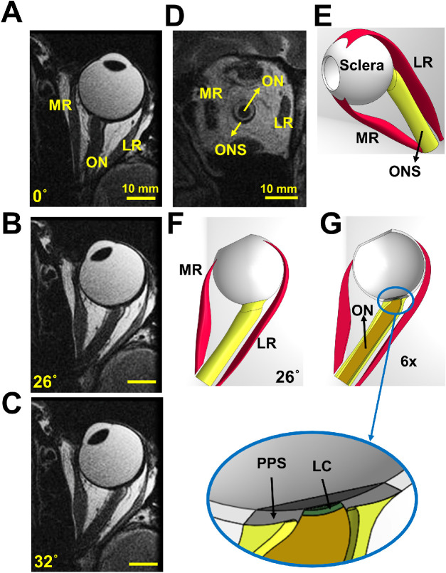
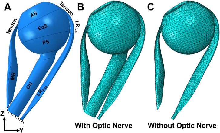
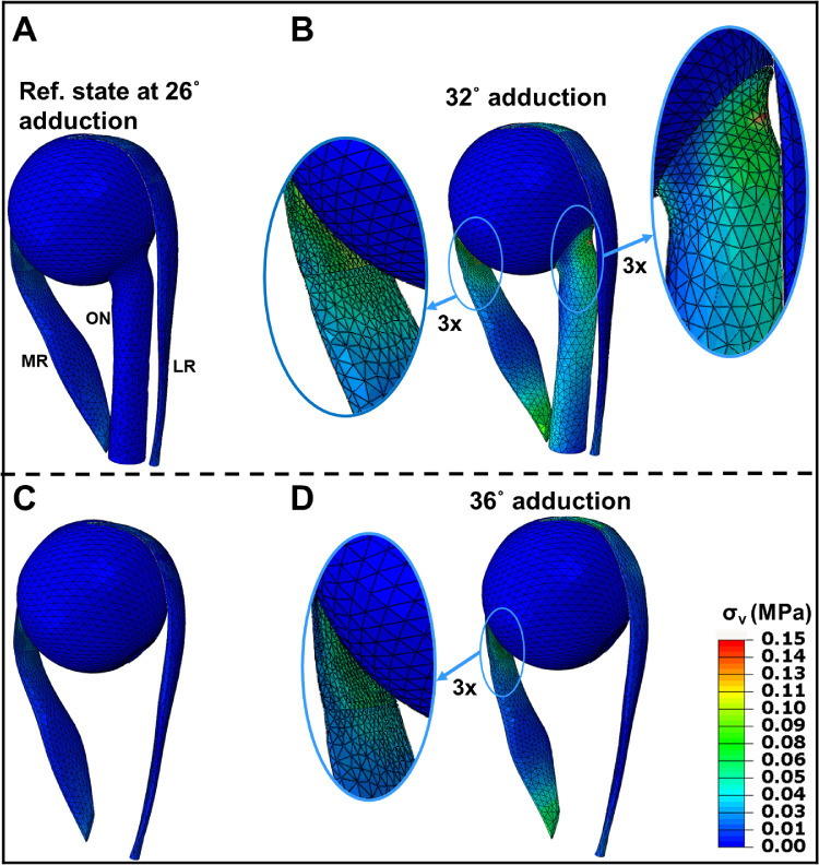
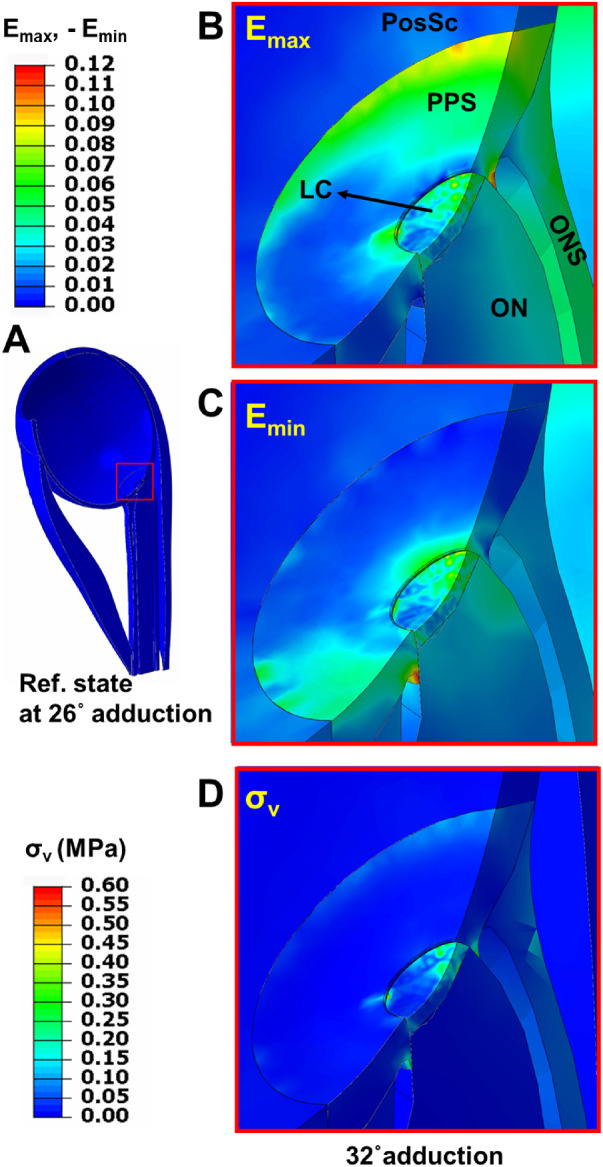
Three-dimensional (3-D) horizontal rectus EOM geometries were obtained from magnetic resonance imaging of five healthy adults, and measured constitutive tissue properties were used. Active and passive strain energies of EOMs were defined using ABAQUS (Dassault Systemes) software. All deformations were assumed to be caused by EOM twitch activation that rotated the eye about a fixed center. The medial rectus (MR) muscle was commanded to additionally contract starting from 26 degrees adducted position, and the lateral rectus (LR) to relax, further adducting the eye either with or without loading by the ON. Tridimensional heat maps were generated to represent the stress and strain distributions.

RESULTS

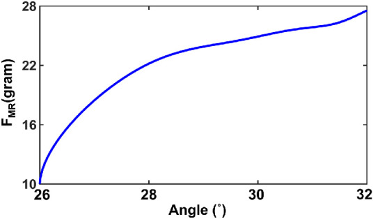
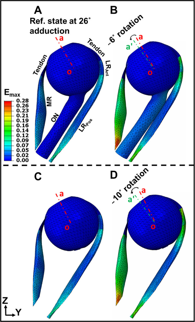
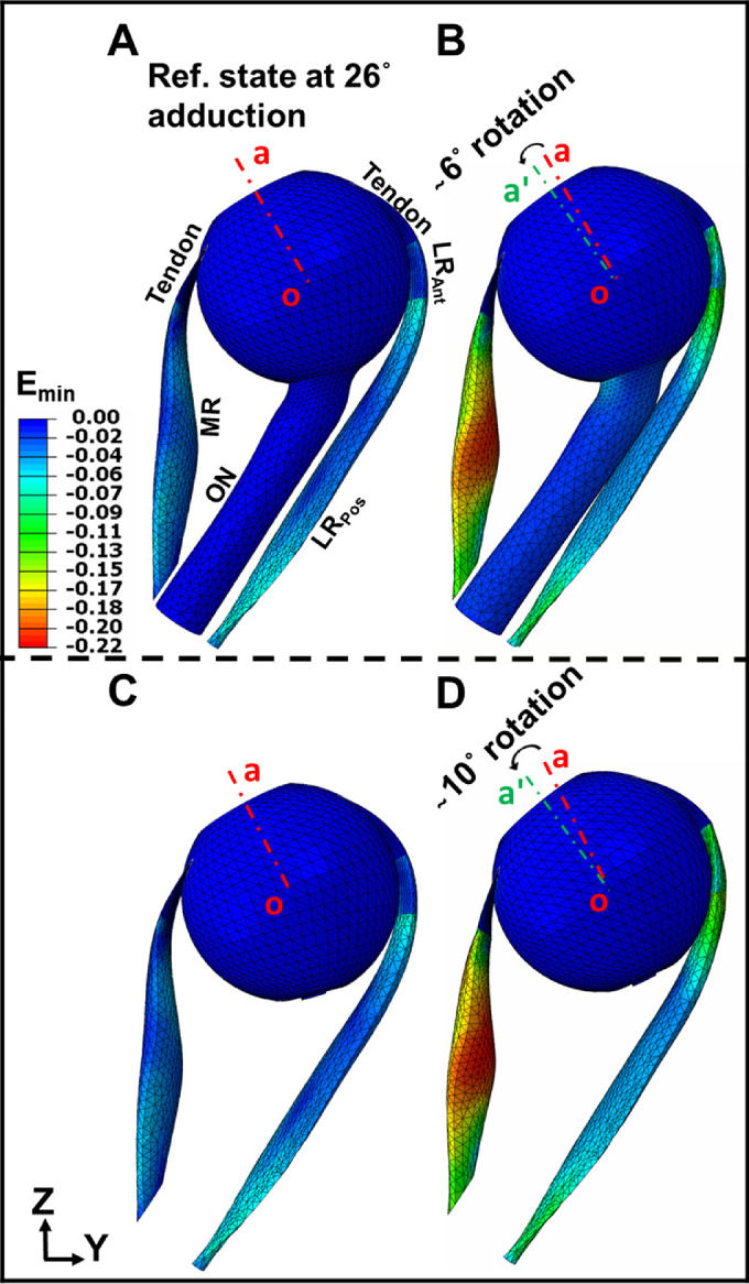
Tensions in the EOMs were physiologically plausible during incremental adduction. Force in the MR increased from 10 gm at 26 degrees adduction to approximately 28 gm at 32 degrees adduction. Under identical MR contraction, adduction with ON loading reached 32 degrees but 36 degrees without it. Maximum and minimum principal strains within the MR were 16% and 22%, respectively, but when ON loading was included, resulting stress and strain were concentrated at the optic disc.

CONCLUSIONS

This physiologically plausible method of simulating EOM activation can provide realistic input to model biomechanical behavior of active and passive tissues in the orbit to clarify biomechanical consequences of ON traction during adduction.

摘要

目的

为了阐明视神经(ON)作为眼球旋转负荷的作用,我们开发了一种有限元模型(FEM),用于模拟主动收缩眼外肌(EOM)引起的增量内收,同时考虑和不考虑 ON 的束缚。

方法

从 5 位健康成年人的磁共振成像中获取了三维(3-D)水平直肌 EOM 几何形状,并使用了测量得到的组织本构特性。使用 ABAQUS(达索系统)软件定义了 EOM 的主动和被动应变能。所有变形都假定是由 EOM 抽搐激活引起的,该激活使眼睛围绕固定中心旋转。命令内侧直肌(MR)在 26 度内收位置进一步收缩,而外侧直肌(LR)放松,使眼睛进一步内收,同时或不加载 ON。生成三维热图来表示应力和应变分布。

结果

在增量内收过程中,EOM 中的张力在生理上是合理的。MR 中的力从 26 度内收时的 10 克增加到 32 度内收时的约 28 克。在相同的 MR 收缩下,加载 ON 时可以达到 32 度内收,但没有 ON 加载时只能达到 36 度内收。MR 中的最大和最小主应变分别为 16%和 22%,但当包括 ON 加载时,导致的应力和应变集中在视盘处。

结论

这种模拟 EOM 激活的生理上合理的方法可以为模型的轨道中主动和被动组织的生物力学行为提供现实的输入,以阐明内收过程中 ON 牵引的生物力学后果。

https://cdn.ncbi.nlm.nih.gov/pmc/blobs/2ea9/7794280/afd2ea7ee8cf/iovs-62-1-1-f010.jpg
https://cdn.ncbi.nlm.nih.gov/pmc/blobs/2ea9/7794280/da532d70226d/iovs-62-1-1-f001.jpg
https://cdn.ncbi.nlm.nih.gov/pmc/blobs/2ea9/7794280/add4938bbf2c/iovs-62-1-1-f002.jpg
https://cdn.ncbi.nlm.nih.gov/pmc/blobs/2ea9/7794280/eb6e37116d85/iovs-62-1-1-f003.jpg
https://cdn.ncbi.nlm.nih.gov/pmc/blobs/2ea9/7794280/a2bef609484f/iovs-62-1-1-f004.jpg
https://cdn.ncbi.nlm.nih.gov/pmc/blobs/2ea9/7794280/bf415783e438/iovs-62-1-1-f005.jpg
https://cdn.ncbi.nlm.nih.gov/pmc/blobs/2ea9/7794280/977ec95dbaa0/iovs-62-1-1-f006.jpg
https://cdn.ncbi.nlm.nih.gov/pmc/blobs/2ea9/7794280/98b4577c5978/iovs-62-1-1-f007.jpg
https://cdn.ncbi.nlm.nih.gov/pmc/blobs/2ea9/7794280/e8742f3534ad/iovs-62-1-1-f008.jpg
https://cdn.ncbi.nlm.nih.gov/pmc/blobs/2ea9/7794280/48b9a03c75d0/iovs-62-1-1-f009.jpg
https://cdn.ncbi.nlm.nih.gov/pmc/blobs/2ea9/7794280/afd2ea7ee8cf/iovs-62-1-1-f010.jpg

相似文献

1
Finite Element Model of Ocular Adduction by Active Extraocular Muscle Contraction.主动眼外肌收缩的眼球内收的有限元模型。
Invest Ophthalmol Vis Sci. 2021 Jan 4;62(1):1. doi: 10.1167/iovs.62.1.1.
2
Finite element model of ocular adduction with unconstrained globe translation.眼球内收的无约束眼球平移有限元模型。
Biomech Model Mechanobiol. 2024 Apr;23(2):601-614. doi: 10.1007/s10237-023-01794-3. Epub 2024 Feb 28.
3
Non-commutative, nonlinear, and non-analytic aspects of the ocular motor plant.眼运动器官的非交换、非线性和非解析方面。
Prog Brain Res. 2019;248:93-102. doi: 10.1016/bs.pbr.2019.04.015. Epub 2019 May 20.
4
Magnetic resonance imaging of differential compartmental function of horizontal rectus extraocular muscles during conjugate and converged ocular adduction.水平直肌在共轭性和集合性眼球内收过程中不同肌间隔功能的磁共振成像
J Neurophysiol. 2014 Aug 15;112(4):845-55. doi: 10.1152/jn.00649.2013. Epub 2014 May 21.
5
Functional morphometry of horizontal rectus extraocular muscles during horizontal ocular duction.水平眼球引出时水平直肌眼外肌的功能形态测量。
Invest Ophthalmol Vis Sci. 2012 Oct 25;53(11):7375-9. doi: 10.1167/iovs.12-9730.
6
Changes in Extraocular Muscle Volume During Ocular Duction.眼球运动时眼外肌体积的变化
Invest Ophthalmol Vis Sci. 2016 Mar;57(3):1106-11. doi: 10.1167/iovs.15-18705.
7
Magnetic Resonance Imaging of the Globe-Tendon Interface for Extraocular Muscles: Is There an "Arc of Contact"?眼球外肌肌腱-球结合部的磁共振成像:是否存在“接触弧”?
Am J Ophthalmol. 2018 Oct;194:170-181. doi: 10.1016/j.ajo.2018.07.002. Epub 2018 Jul 19.
8
Extraocular muscle deformation assessed by motion-encoded MRI during eye movement in healthy subjects.健康受试者眼球运动期间通过运动编码MRI评估眼外肌变形
J Vis. 2007 Nov 21;7(14):5.1-10. doi: 10.1167/7.14.5.
9
Predictions of Optic Nerve Traction Forces and Peripapillary Tissue Stresses Following Horizontal Eye Movements.水平眼球运动后视神经牵引力和视乳头周围组织应力的预测
Invest Ophthalmol Vis Sci. 2017 Apr 1;58(4):2044-2053. doi: 10.1167/iovs.16-21319.
10
Local deformation of extraocular muscles during eye movement.眼球运动过程中眼外肌的局部变形。
Invest Ophthalmol Vis Sci. 2009 Nov;50(11):5189-96. doi: 10.1167/iovs.08-3182. Epub 2009 Jun 10.

引用本文的文献

1
Biomechanical Analysis and Clinical Study of Augmented Versus Conventional Endoscopic Orbital Decompression for Dysthyroid Optic Neuropathy.甲状腺相关性视神经病变的增强型与传统内镜下眼眶减压术的生物力学分析及临床研究
Bioengineering (Basel). 2025 Jun 5;12(6):618. doi: 10.3390/bioengineering12060618.
2
Shear viscoelastic properties of human orbital fat.人体眼眶脂肪的剪切粘弹性特性
J Biomech. 2024 Dec;177:112416. doi: 10.1016/j.jbiomech.2024.112416. Epub 2024 Nov 19.
3
Tensile properties of glaucomatous human sclera, optic nerve, and optic nerve sheath.

本文引用的文献

1
Adduction-Induced Strain on the Optic Nerve in Primary Open Angle Glaucoma at Normal Intraocular Pressure.正常眼压原发性开角型青光眼视神经营养不良时的内收诱导性视神经应变。
Curr Eye Res. 2021 Apr;46(4):568-578. doi: 10.1080/02713683.2020.1817491. Epub 2020 Sep 11.
2
Compression of the Choroid by Horizontal Duction.脉络膜受压导致水平牵引。
Invest Ophthalmol Vis Sci. 2019 Oct 1;60(13):4285-4291. doi: 10.1167/iovs.19-27522.
3
Age-dependent Deformation of the Optic Nerve Head and Peripapillary Retina by Horizontal Duction.水平牵引对视盘和视盘周围视网膜的年龄相关性变形作用
青光眼患者巩膜、视神经和视神经鞘的拉伸性能。
Biomech Model Mechanobiol. 2024 Dec;23(6):1851-1862. doi: 10.1007/s10237-024-01872-0. Epub 2024 Aug 8.
4
Postmortem Digital Image Correlation and Finite Element Modeling Demonstrate Posterior Scleral Deformations during Optic Nerve Adduction Tethering.尸检数字图像相关分析和有限元建模显示视神经内收束缚期间后巩膜变形。
Bioengineering (Basel). 2024 May 2;11(5):452. doi: 10.3390/bioengineering11050452.
5
Ciliary muscles contraction leads to axial length extension--The possible initiating factor for myopia.睫状肌收缩导致眼轴延长——近视的可能起始因素。
PLoS One. 2024 Apr 16;19(4):e0301844. doi: 10.1371/journal.pone.0301844. eCollection 2024.
6
Scanning Laser Ophthalmoscopy Demonstrates Pediatric Optic Disc and Peripapillary Strain During Horizontal Eye Rotation.扫描激光检眼镜显示儿童视盘和视盘周围在眼球水平转动时的应变。
Curr Eye Res. 2024 Apr;49(4):437-445. doi: 10.1080/02713683.2023.2295789. Epub 2024 Jan 7.
7
Empirical Quantification of Optic Nerve Strain Due to Horizontal Duction.水平眼外肌运动引起的视神经应变的实证量化
Bioengineering (Basel). 2023 Aug 5;10(8):931. doi: 10.3390/bioengineering10080931.
8
Scanning Laser Ophthalmoscopy Demonstrates Disc and Peripapillary Strain During Horizontal Eye Rotation in Adults.扫描激光检眼镜显示成人眼球水平转动时视盘和视盘周围的应变。
Am J Ophthalmol. 2023 Oct;254:114-127. doi: 10.1016/j.ajo.2023.06.008. Epub 2023 Jun 19.
9
Who bears the load? IOP-induced collagen fiber recruitment over the corneoscleral shell.谁来承担这个负担?IOP 诱导的角膜巩膜壳上的胶原纤维募集。
Exp Eye Res. 2023 May;230:109446. doi: 10.1016/j.exer.2023.109446. Epub 2023 Mar 18.
10
Linear viscoelasticity of human sclera and posterior ocular tissues during tensile creep.人眼球巩膜和眼后组织在拉伸蠕变过程中的线性粘弹性。
J Biomech. 2023 Apr;151:111530. doi: 10.1016/j.jbiomech.2023.111530. Epub 2023 Mar 11.
Am J Ophthalmol. 2020 Jan;209:107-116. doi: 10.1016/j.ajo.2019.08.017. Epub 2019 Aug 29.
4
Optic Nerve Traction During Adduction in Open Angle Glaucoma with Normal versus Elevated Intraocular Pressure.在正常眼压和高眼压的开角型青光眼患者中,眼球内收时视神经的牵拉。
Curr Eye Res. 2020 Feb;45(2):199-210. doi: 10.1080/02713683.2019.1660371. Epub 2019 Dec 2.
5
Translation and eccentric rotation in ocular motor modeling.眼球运动建模中的翻译和偏旋。
Prog Brain Res. 2019;248:117-126. doi: 10.1016/bs.pbr.2019.04.036. Epub 2019 May 23.
6
Rectus Extraocular Muscle Paths and Staphylomata in High Myopia.高度近视患者直肌眼外肌路径和眼球突出。
Am J Ophthalmol. 2019 May;201:37-45. doi: 10.1016/j.ajo.2019.01.029. Epub 2019 Feb 4.
7
Adduction-Induced Deformations Evoke Peripapillary Folds in Papilledema.内收诱发的变形引发视乳头水肿中的视乳头周围褶皱。
Ophthalmology. 2019 Jun;126(6):912-914. doi: 10.1016/j.ophtha.2018.12.043. Epub 2018 Dec 24.
8
Optic Nerve Sheath Tethering in Adduction Occurs in Esotropia and Hypertropia, But Not in Exotropia.内收时视神经鞘牵拉仅见于内斜视和上斜视,而外斜视则不然。
Invest Ophthalmol Vis Sci. 2018 Jun 1;59(7):2899-2904. doi: 10.1167/iovs.18-24305.
9
Progressive Deformation of the Optic Nerve Head and Peripapillary Structures by Graded Horizontal Duction.通过分级水平转位对视神经乳头和视乳头周围结构进行渐进性变形
Invest Ophthalmol Vis Sci. 2017 Oct 1;58(12):5015-5021. doi: 10.1167/iovs.17-22596.
10
Prediction of muscle activation for an eye movement with finite element modeling.利用有限元模型预测眼球运动中的肌肉激活情况。
Comput Biol Med. 2017 Oct 1;89:368-378. doi: 10.1016/j.compbiomed.2017.08.018. Epub 2017 Aug 24.