• 文献检索
  • 文档翻译
  • 深度研究
  • 学术资讯
  • Suppr Zotero 插件Zotero 插件
  • 邀请有礼
  • 套餐&价格
  • 历史记录
应用&插件
Suppr Zotero 插件Zotero 插件浏览器插件Mac 客户端Windows 客户端微信小程序
定价
高级版会员购买积分包购买API积分包
服务
文献检索文档翻译深度研究API 文档MCP 服务
关于我们
关于 Suppr公司介绍联系我们用户协议隐私条款
关注我们

Suppr 超能文献

核心技术专利:CN118964589B侵权必究
粤ICP备2023148730 号-1Suppr @ 2026

文献检索

告别复杂PubMed语法,用中文像聊天一样搜索,搜遍4000万医学文献。AI智能推荐,让科研检索更轻松。

立即免费搜索

文件翻译

保留排版,准确专业,支持PDF/Word/PPT等文件格式,支持 12+语言互译。

免费翻译文档

深度研究

AI帮你快速写综述,25分钟生成高质量综述,智能提取关键信息,辅助科研写作。

立即免费体验

胸骨切开术后 F-FDG/PET-CT 影像学表现。

F-FDG/PET-CT imaging findings after sternotomy.

机构信息

Department of Radiology and Nuclear Medicine, Erasmus Medical Center, ND-547, Dr. Molewaterplein 40, 015GD, Rotterdam, The Netherlands.

Department of Cardiology, Thoraxcenter, Erasmus Medical Center, Rotterdam, The Netherlands.

出版信息

J Nucl Cardiol. 2023 Jun;30(3):1210-1218. doi: 10.1007/s12350-022-03126-x. Epub 2022 Nov 8.

DOI:10.1007/s12350-022-03126-x
PMID:36348248
原文链接:https://pmc.ncbi.nlm.nih.gov/articles/PMC10261398/
Abstract

BACKGROUND

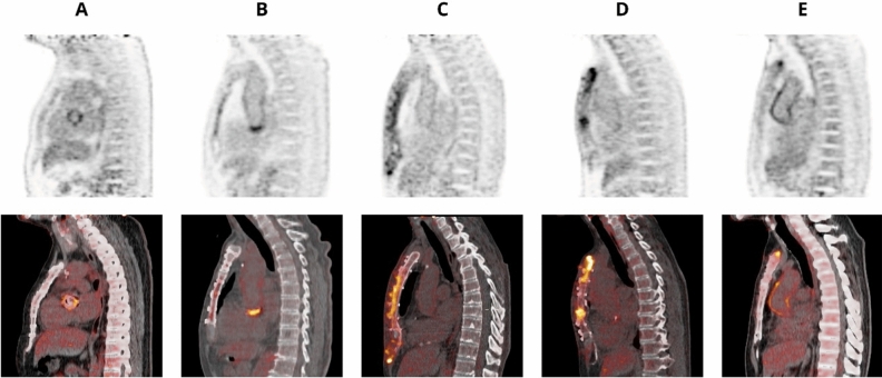
The clinical diagnosis of deep sternal wound infection (DSWI) is supported by imaging findings including 18F-fluorodeoxyglucose positron emission tomography/computed tomography (F-FDG-PET/CT). To avoid misinterpretation due to normal post-surgery inflammation we assessed normal imaging findings in non-infected patients after sternotomy.

METHODS

This is a prospective cohort study including non-infectious patients with sternotomy. All patients underwent F-FDG-PET/CT at either 5 weeks (group 1), 12 weeks (group 2) or 52 weeks (group 3) post-surgery. F-FDG uptake was scored visually in five categories and assessed quantitatively.

RESULTS

A total of 44 patients were included. Sternal mean SUVmax was 7.34 (± 1.86), 5.22 (± 2.55) and 3.20 (± 1.80) in group 1, 2 and 3, respectively (p < 0.01). Sternal mean SUVmean was 3.84 (± 1.00), 2.69 (± 1.32) and 1.71 (± 0.98) in group 1, 2 and 3 (p < 0.01). All patients in group 1 had elevated uptake whereas group 2 and 3 showed 2/15 (13%) and 11/20 (55%) patients respectively with no elevated uptake. Group 3 still showed an elevated uptake pattern in in 9/20 (45%) and in 3/9 (33%) with a high-grade diffuse uptake pattern.

CONCLUSION

This study shows significant lower sternal F-FDG at 55 weeks compared to 5 weeks post-sternotomy however elevated uptake patterns may persist.

摘要

背景

18F-氟代脱氧葡萄糖正电子发射断层扫描/计算机断层扫描(F-FDG-PET/CT)的影像学表现支持深部胸骨伤口感染(DSWI)的临床诊断。为了避免由于手术后正常炎症而导致的误诊,我们评估了胸骨切开术后非感染患者的正常影像学表现。

方法

这是一项包括非感染性胸骨切开术患者的前瞻性队列研究。所有患者均在手术后 5 周(第 1 组)、12 周(第 2 组)或 52 周(第 3 组)进行 F-FDG-PET/CT 检查。通过 5 个类别进行视觉评分和定量评估 F-FDG 摄取。

结果

共纳入 44 例患者。第 1、2 和 3 组胸骨 SUVmax 的平均值分别为 7.34(±1.86)、5.22(±2.55)和 3.20(±1.80)(p<0.01)。第 1、2 和 3 组胸骨 SUVmean 的平均值分别为 3.84(±1.00)、2.69(±1.32)和 1.71(±0.98)(p<0.01)。第 1 组所有患者摄取均升高,而第 2 和第 3 组分别有 2/15(13%)和 11/20(55%)的患者摄取不升高。第 3 组在 20 例患者中有 9 例(45%)和在 9 例中有 3 例(33%)仍表现为摄取升高模式,表现为高分级弥漫性摄取模式。

结论

本研究表明,与胸骨切开术后 5 周相比,55 周时胸骨 F-FDG 摄取显著降低,但摄取升高模式可能持续存在。

https://cdn.ncbi.nlm.nih.gov/pmc/blobs/bf0a/10261398/3bdbf15bf45f/12350_2022_3126_Fig3_HTML.jpg
https://cdn.ncbi.nlm.nih.gov/pmc/blobs/bf0a/10261398/f0e3dedcfc41/12350_2022_3126_Fig1_HTML.jpg
https://cdn.ncbi.nlm.nih.gov/pmc/blobs/bf0a/10261398/7edb0f8d3eb1/12350_2022_3126_Fig2_HTML.jpg
https://cdn.ncbi.nlm.nih.gov/pmc/blobs/bf0a/10261398/3bdbf15bf45f/12350_2022_3126_Fig3_HTML.jpg
https://cdn.ncbi.nlm.nih.gov/pmc/blobs/bf0a/10261398/f0e3dedcfc41/12350_2022_3126_Fig1_HTML.jpg
https://cdn.ncbi.nlm.nih.gov/pmc/blobs/bf0a/10261398/7edb0f8d3eb1/12350_2022_3126_Fig2_HTML.jpg
https://cdn.ncbi.nlm.nih.gov/pmc/blobs/bf0a/10261398/3bdbf15bf45f/12350_2022_3126_Fig3_HTML.jpg

相似文献

1
F-FDG/PET-CT imaging findings after sternotomy.胸骨切开术后 F-FDG/PET-CT 影像学表现。
J Nucl Cardiol. 2023 Jun;30(3):1210-1218. doi: 10.1007/s12350-022-03126-x. Epub 2022 Nov 8.
2
More advantages in detecting bone and soft tissue metastases from prostate cancer using F-PSMA PET/CT.使用F-PSMA PET/CT检测前列腺癌骨和软组织转移方面有更多优势。
Hell J Nucl Med. 2019 Jan-Apr;22(1):6-9. doi: 10.1967/s002449910952. Epub 2019 Mar 7.
3
Diagnostic value of fluorine-18 deoxyglucose positron emission tomography/computed tomography in deep sternal wound infection.氟-18脱氧葡萄糖正电子发射断层扫描/计算机断层扫描在胸骨深部伤口感染中的诊断价值
J Plast Reconstr Aesthet Surg. 2018 Dec;71(12):1768-1776. doi: 10.1016/j.bjps.2018.07.017. Epub 2018 Aug 2.
4
Prone position PET/CT is useful in reducing gravity-dependent opacity-related [F]fluorodeoxyglucose uptake.俯卧位 PET/CT 有助于减少与重力相关的不透明性有关的 [F]氟脱氧葡萄糖摄取。
Eur Radiol. 2023 Nov;33(11):8270-8278. doi: 10.1007/s00330-023-09831-6. Epub 2023 Jun 16.
5
18F-Fluorodeoxyglucose Positron Emission Tomography/Magnetic Resonance in Lymphoma: Comparison With 18F-Fluorodeoxyglucose Positron Emission Tomography/Computed Tomography and With the Addition of Magnetic Resonance Diffusion-Weighted Imaging.18F-氟脱氧葡萄糖正电子发射断层扫描/磁共振成像在淋巴瘤中的应用:与18F-氟脱氧葡萄糖正电子发射断层扫描/计算机断层扫描的比较以及添加磁共振扩散加权成像的研究
Invest Radiol. 2016 Mar;51(3):163-9. doi: 10.1097/RLI.0000000000000218.
6
Appearance of aseptic vascular grafts after endovascular aortic repair on [(18)F]fluorodeoxyglucose positron emission tomography/computed tomography.经皮腔内主动脉修复术后无菌血管移植物在[(18)F]氟脱氧葡萄糖正电子发射断层扫描/计算机断层扫描上的表现
World J Radiol. 2023 Aug 28;15(8):241-249. doi: 10.4329/wjr.v15.i8.241.
7
The Diagnostic Value of Fluorine-18 Fluorodeoxyglucose Positron Emission Tomography/Computed Tomography for the Detection of Surgical Site Infection after Spine Surgery.氟-18氟脱氧葡萄糖正电子发射断层扫描/计算机断层扫描在脊柱手术后手术部位感染检测中的诊断价值
Spine (Phila Pa 1976). 2021 May 15;46(10):E602-E610. doi: 10.1097/BRS.0000000000003847.
8
The value of F-FDG PET/CT in diagnosing and localising deep sternal wound infection to guide surgical debridement.F-FDG PET/CT 在诊断和定位深部胸骨伤口感染以指导外科清创中的价值。
Int Wound J. 2020 Aug;17(4):1019-1027. doi: 10.1111/iwj.13368. Epub 2020 Apr 16.
9
Comparison of [F]fluciclovine and [F]FDG PET/CT in Newly Diagnosed Multiple Myeloma Patients.新型多发性骨髓瘤患者中[F]氟替卡松和[F]FDG PET/CT 的比较。
Mol Imaging Biol. 2022 Oct;24(5):842-851. doi: 10.1007/s11307-022-01734-0. Epub 2022 May 2.
10
[F]FDG PET/CT in non-union: improving the diagnostic performances by using both PET and CT criteria.[F]FDG PET/CT 在骨不连中的应用:同时使用 PET 和 CT 标准提高诊断性能。
Eur J Nucl Med Mol Imaging. 2019 Jul;46(8):1605-1615. doi: 10.1007/s00259-019-04336-1. Epub 2019 May 1.

引用本文的文献

1
Prospective evaluation of the diagnostic accuracy of FDG-PET/CT for the detection of sternal wound infection post-sternotomy.18F-氟脱氧葡萄糖正电子发射断层扫描/计算机断层扫描(FDG-PET/CT)检测胸骨切开术后胸骨伤口感染诊断准确性的前瞻性评估。
EJNMMI Rep. 2025 Feb 8;9(1):6. doi: 10.1186/s41824-024-00237-1.

本文引用的文献

1
Normal imaging findings after ascending aorta prosthesis implantation on F-Fluorodeoxyglucose Positron Emission Tomography with computed tomography.升主动脉假体植入后 F-氟脱氧葡萄糖正电子发射断层扫描与计算机断层扫描的正常影像学表现。
J Nucl Cardiol. 2022 Dec;29(6):2938-2948. doi: 10.1007/s12350-021-02826-0. Epub 2021 Oct 27.
2
The value of F-FDG PET/CT in diagnosing and localising deep sternal wound infection to guide surgical debridement.F-FDG PET/CT 在诊断和定位深部胸骨伤口感染以指导外科清创中的价值。
Int Wound J. 2020 Aug;17(4):1019-1027. doi: 10.1111/iwj.13368. Epub 2020 Apr 16.
3
Normal imaging findings after aortic valve implantation on F-Fluorodeoxyglucose positron emission tomography with computed tomography.
经 F-Fluorodeoxyglucose 正电子发射断层扫描与计算机断层扫描的主动脉瓣植入术后正常影像学表现。
J Nucl Cardiol. 2021 Oct;28(5):2258-2268. doi: 10.1007/s12350-019-02025-y. Epub 2020 Jan 23.
4
Utility of FDG-PET/CT for the Detection and Characterization of Sternal Wound Infection Following Sternotomy.18F-氟脱氧葡萄糖正电子发射断层显像/计算机断层扫描(FDG-PET/CT)在胸骨切开术后胸骨伤口感染检测及特征分析中的应用
Nucl Med Mol Imaging. 2019 Aug;53(4):253-262. doi: 10.1007/s13139-019-00599-6. Epub 2019 Jun 14.
5
Diagnostic value of fluorine-18 deoxyglucose positron emission tomography/computed tomography in deep sternal wound infection.氟-18脱氧葡萄糖正电子发射断层扫描/计算机断层扫描在胸骨深部伤口感染中的诊断价值
J Plast Reconstr Aesthet Surg. 2018 Dec;71(12):1768-1776. doi: 10.1016/j.bjps.2018.07.017. Epub 2018 Aug 2.
6
Prevention and management of sternal wound infections.胸骨伤口感染的预防与管理
J Thorac Cardiovasc Surg. 2016 Oct;152(4):962-72. doi: 10.1016/j.jtcvs.2016.01.060. Epub 2016 Aug 8.
7
PET-CT imaging in patients with chronic sternal wound infections prior to reconstructive surgery: A case series.慢性胸骨伤口感染患者重建手术前的PET-CT成像:病例系列
J Plast Reconstr Aesthet Surg. 2015 Aug;68(8):1132-7. doi: 10.1016/j.bjps.2015.04.027. Epub 2015 May 7.
8
Diagnostic value of ⁹⁹mTc-HMPAO-labeled leukocytes scintigraphy in suspicion of post-sternotomy mediastinitis relapse.⁹⁹mTc-HMPAO 标记白细胞闪烁扫描术对怀疑胸骨切开术后纵隔炎复发的诊断价值
J Nucl Cardiol. 2015 Feb;22(1):123-9. doi: 10.1007/s12350-014-9999-9. Epub 2014 Oct 8.
9
EANM/SNMMI guideline for 18F-FDG use in inflammation and infection.欧洲核医学协会/美国核医学与分子影像学会18F-FDG在炎症和感染中应用的指南
J Nucl Med. 2013 Apr;54(4):647-58. doi: 10.2967/jnumed.112.112524. Epub 2013 Jan 28.
10
Epidemiology of deep sternal wound infection in cardiac surgery.心脏手术中深部胸骨伤口感染的流行病学
J Cardiothorac Vasc Anesth. 2009 Aug;23(4):488-94. doi: 10.1053/j.jvca.2009.02.007. Epub 2009 Apr 19.