Giammalva Giuseppe Roberto, Viola Anna, Maugeri Rosario, Giardina Kevin, Di Bonaventura Rina, Musso Sofia, Brunasso Lara, Cepeda Santiago, Della Pepa Giuseppe Maria, Scerrati Alba, Mantovani Giorgio, Ferini Gianluca, Gerardi Rosa Maria, Pino Maria Angela, Umana Giuseppe Emmanuele, Denaro Luca, Albanese Alessio, Iacopino Domenico Gerardo
Neurosurgical Clinic, Post Graduate Residency Program in Neurologic Surgery, Department of Biomedicine Neurosciences and Advanced Diagnostics, School of Medicine, University of Palermo, 90127 Palermo, Italy.
Department of Radiation Oncology, REM Radioterapia srl, 95029 Viagrande, Italy.
Cancers (Basel). 2022 Oct 29;14(21):5335. doi: 10.3390/cancers14215335.
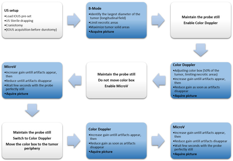
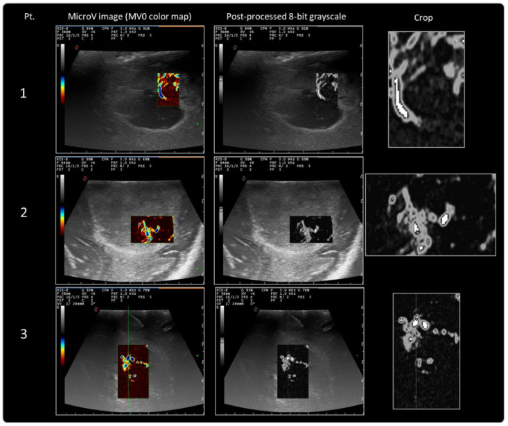
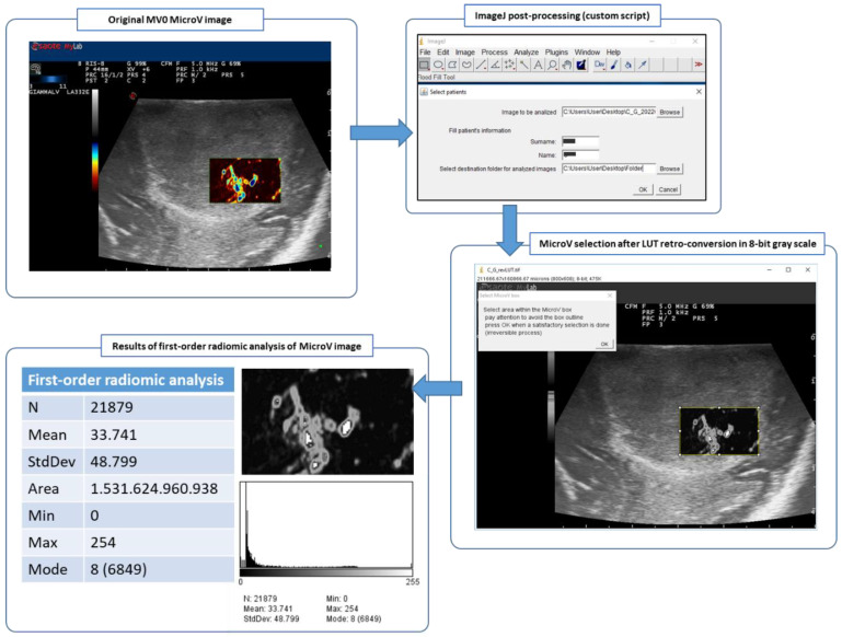
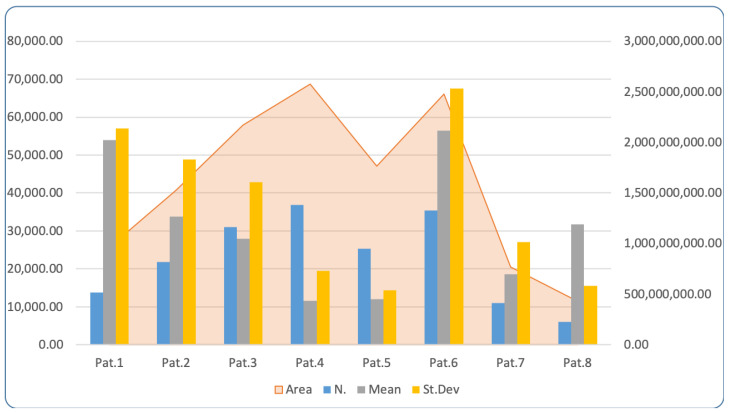
Microvascular Doppler (MicroV) is a new-generation Doppler technique developed by Esaote (Esaote s.p.a., Genova, Italy), which is able to visualize small and low-flow vessels through a suppression of interfering signals. MicroV uses advanced filters that are able to differentiate tissue artifacts from low-speed blood flows; by exploiting the space-time coherence information, these filters can selectively suppress tissue components, preserving the signal coming from the microvascular flow. This technique is clinically applied to the study of the vascularization of parenchymatous lesions, often with better diagnostic accuracy than color/power Doppler techniques. The aim of this paper is to develop a reproducible protocol for the recording and collection of MicroV intraoperative ultrasound images by the use of a capable intraoperative ultrasound machine and post-processing aimed at evaluation of brain-tumor microvascularization through the analysis of radiomic features. The proposed protocol has been internally validated on eight patients and will be firstly applied to patients affected by WHO grade IV astrocytoma (glioblastoma-GBM) candidates for craniotomy and lesion removal. In a further stage, it will be generally applied to patients with primary or metastatic brain tumors. IOUS is performed before durotomy. Tumor microvascularization is evaluated using the MicroV Doppler technique and IOUS images are recorded, stored, and post-processed. IOUS images are remotely stored on the BraTIoUS database, which will promote international cooperation and multicentric analysis. Processed images and texture radiomic features are analyzed post-operatively using ImageJ, a free scientific image-analysis software based on the Sun-Java platform. Post-processing protocol is further described in-depth. The study of tumor microvascularization through advanced IOUS techniques such as MicroV could represent, in the future, a non-invasive and real-time method for intraoperative predictive evaluation of the tumor features. This evaluation could finally result in a deeper knowledge of brain-tumor behavior and in the on-going adaptation of the surgery with the improvement of surgical outcomes.
微血管多普勒(MicroV)是由意大利热那亚的百胜医疗集团(Esaote s.p.a.)研发的新一代多普勒技术,它能够通过抑制干扰信号来可视化小血管和低流量血管。MicroV使用先进的滤波器,能够区分组织伪像和低速血流;通过利用时空相干信息,这些滤波器可以选择性地抑制组织成分,保留来自微血管血流的信号。该技术在临床上用于实质病变血管化的研究,其诊断准确性通常优于彩色/能量多普勒技术。本文的目的是通过使用一台功能强大的术中超声机器以及旨在通过分析影像组学特征评估脑肿瘤微血管化的后处理,制定一种可重复的记录和采集MicroV术中超声图像的方案。所提出的方案已在8名患者身上进行了内部验证,并将首先应用于拟行开颅手术和病变切除的世界卫生组织IV级星形细胞瘤(胶质母细胞瘤-GBM)患者。在进一步的阶段,它将普遍应用于原发性或转移性脑肿瘤患者。在硬脑膜切开术前进行术中超声检查。使用MicroV多普勒技术评估肿瘤微血管化,并记录、存储和后处理术中超声图像。术中超声图像远程存储在BraTIoUS数据库中,这将促进国际合作和多中心分析。术后使用基于Sun-Java平台的免费科学图像分析软件ImageJ分析处理后的图像和纹理影像组学特征。后处理方案将进一步深入描述。通过诸如MicroV等先进的术中超声技术研究肿瘤微血管化,未来可能代表一种用于术中预测评估肿瘤特征的非侵入性实时方法。这种评估最终可能会更深入地了解脑肿瘤的行为,并随着手术结果的改善不断调整手术方式。